. 2025 Jul 8:6:1612522.
doi: 10.3389/fdmed.2025.1612522. eCollection 2025.

Salivary gland transcriptomic analysis and immunophenotyping in the IL-14α transgenic mouse model of Sjögren's disease

Affiliations

Salivary gland transcriptomic analysis and immunophenotyping in the IL-14α transgenic mouse model of Sjögren's disease

Lucas T Woods et al. Front Dent Med. .

Abstract

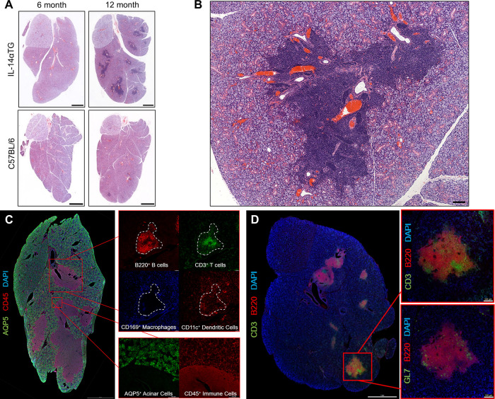
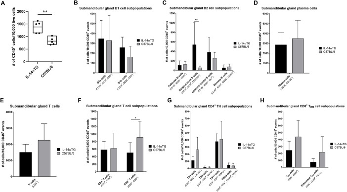
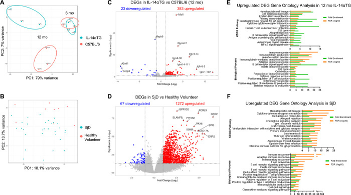
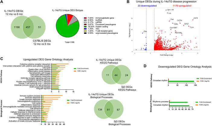
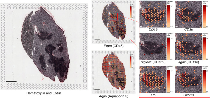
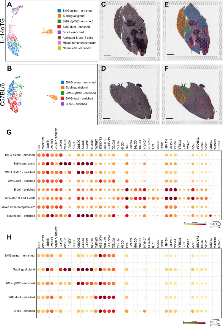
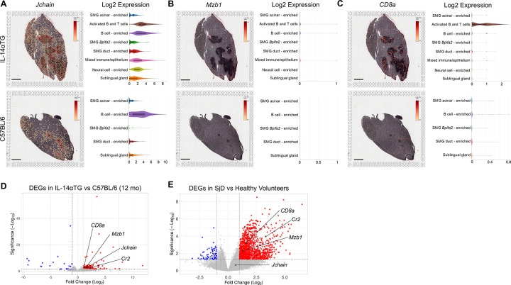
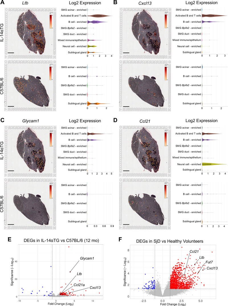
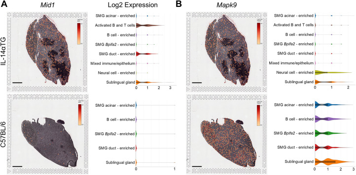
Sjögren's disease (SjD) is a systemic autoimmune disorder primarily affecting the exocrine glands and characterized by dry mouth and dry eye, the presence of anti-SSA and/or anti-SSB autoantibodies in blood serum, and chronic lymphocytic infiltration of salivary and lacrimal glands (i.e., sialadenitis and dacryoadenitis, respectively). In addition to reduced quality of life, SjD patients experience severe oral health complications and are at increased risk of developing B cell lymphoma. Because current SjD treatments primarily focus on oral and ocular symptom management, identifying initiating factors and mechanisms of disease progression may offer new therapeutic insights for SjD. The interleukin-14α transgenic (IL-14αTG) mouse model of SjD recapitulates many aspects of human SjD, including progressive sialadenitis, loss of salivary gland function, and development of B cell lymphoma. We utilized immunofluorescence, flow cytometry, bulk RNA sequencing and spatial transcriptomic analyses to identify immune cell subpopulations and differentially expressed genes (DEGs) in submandibular glands of IL-14αTG Sjögren's-like mice and age-matched C57BL/6 mouse controls. We further compared the gene ontology of DEGs in IL-14αTG mice to DEGs identified in minor salivary gland biopsies from SjD patients and healthy volunteers. Results demonstrated significantly increased sialadenitis in IL-14αTG compared to C57BL/6 mice that correlated with an increased proportion of marginal zone B cells infiltrating the submandibular gland. Whole transcriptome analyses showed substantial overlap in enriched DEG ontology between IL-14αTG mouse submandibular gland and SjD patient minor salivary gland, compared to C57BL/6 mice and healthy human volunteer controls, respectively. Lastly, we spatially resolved DEG expression and localization within IL-14αTG salivary glands, marking the first publication of a spatial transcriptomic dataset from submandibular glands in a SjD mouse model.

Keywords: RNAseq; Sjögren's disease; interleukin-14α transgenic; salivary gland; sialadenitis; spatial transcriptome.


Conflict of interest statement

The authors declare that the research was conducted in the absence of any commercial or financial relationships that could be construed as a potential conflict of interest.

Figures

Figure 1
Figure 1
Progressive sialadenitis in the IL-14αTG mouse model of Sjögren's disease. (A) Submandibular and sublingual glands from 6- and a 12-month-old female IL-14αTG and C57BL/6 mice were subjected to hematoxylin and eosin staining to assess glandular inflammation; scale bar = 1 mm. (B) Immune cell focus surrounding blood vessels and excretory ducts in a 12-month-old IL-14αTG mouse SMG; scale bar = 100 µm. (C) Twelve-month-old and (D) 18-month-old female IL-14αTG mouse SMG and SLG cryosections were subjected to immunofluorescence staining using antibodies against aquaporin 5 (AQP5) acinar cell marker, CD45 pan-immune cell marker, B220 B cell marker, CD3 T cell marker, CD169 macrophage marker, CD11c dendritic cell marker or GL7 germinal center marker with DAPI nuclear counterstain; scale bars = 1 mm and 100 µm (inset).
Figure 2
Figure 2
Flow cytometry analysis of IL-14αTG and C57BL/6 mouse SMG-infiltrating immune cells. SMGs from 12-month-old IL-14αTG and C57BL/6 mice were enzymatically dispersed, stained with either a B or T cell type-specific antibody panel and analyzed by flow cytometry on a Cytek Aurora spectral analyzer. (A) Total CD45+ immune cells, (B–D) B cell subpopulations and (E–H) T cell subpopulations were quantified, and data are presented as means ± S.D., where * and ** indicate P < 0.05 and 0.01, respectively, for n = 5 mice per group.
Figure 3
Figure 3
Comparison of differentially expressed genes from IL-14αTG mouse submandibular glands and human SjD minor salivary glands. RNA prepared from 6- and 12-month-old IL-14αTG and C57BL/6 SMGs was subjected to RNAseq analysis. Previously performed RNAseq analysis of SjD patient and healthy volunteer MSG was accessed through dbGAP accession phs001842.v1.p1. Up- and downregulated DEGs were identified and analyzed by (A,B) principal component analysis, (C,D) DEG volcano plots and (E,F) DAVID gene ontology analysis, where (A,C,E) are results from IL-14αTG vs. C57BL/6 mouse SMGs (n = 3/timepoint/genotype) and (B,D,F) are results from SjD patient vs. healthy volunteer MSG biopsies (n = 35 SjD patients and 9 healthy volunteers).
Figure 4
Figure 4
Unique differentially expressed genes in IL-14αTG mouse submandibular glands. DEG lists from 12-month-old vs. 6-month-old IL-14αTG and 12-month-old vs. 6-month-old C57BL/6 mouse SMGs were compared and genes that were differentially expressed with age in both genotypes were removed to generate a list of DEGs unique to IL-14αTG SMG. (A) Venn diagram of shared and unique genes differentially expressed with age and pie chart of IL-14αTG unique DEG biotype; TEC,To be Experimentally Confirmed. (B) Volcano plot of unique DEGs in IL-14αTG mouse SMG. (C) DAVID gene ontology analysis of upregulated unique IL-14αTG mouse SMG DEGs to identify enriched KEGG pathways and biological processes, with Venn diagrams denoting shared and unique signaling pathway enrichment between IL-14αTG mouse SMG and human SjD MSG biopsies. (D) DAVID gene ontology analysis of downregulated unique IL-14αTG mouse SMG DEGs.
Figure 5
Figure 5
Spatially resolved gene marker expression in IL-14αTG mouse salivary glands using spatial transcriptomic analysis. Fresh frozen SMG and SLG cryosections from a 12-month-old female IL-14αTG mouse were adhered to a barcoded Visium spatial gene expression slide, stained with hematoxylin and eosin, visualized on a Zeiss Axiovert 200M inverted microscope, and then subjected to RNAseq analysis. Color-coded expression of individual gene markers [Log2(counts/capture area)] in barcoded mRNA capture areas was overlaid on the tissue image using Loupe browser 8.1 software. Scale bar = 1 mm and 100 µm (inset).
Figure 6
Figure 6
Spatial transcriptomic analysis and cell clustering of IL-14αTG and C57BL/6 mouse salivary glands. RNAseq analyses of barcoded Visium spatial mRNA capture areas from 12-month-old female IL-14αTG or C57BL/6 mouse SMG and SLG were subjected to unsupervised Louvain clustering and visualization using Bioturing software. (A,B) UMAP and color-coded cell cluster identification, (C,D) hematoxylin and eosin (H&E)-stained tissue images, (E,F) cell cluster spatial localization overlay on H&E tissue images, and (G,H) dot plot of cell cluster-defining gene markers in (A,C,E) IL-14αTG and (B,D,F) C57BL/6 mouse SMG and SLG. Scale bar = 1 mm.
Figure 7
Figure 7
Immune cell gene marker expression, localization, and cell cluster distribution in IL-14αTG and C57BL/6 mouse salivary glands. Color-coded expression [Log2(counts/capture area)] and cell cluster distribution violin plots of (A) Jchain (plasma cell marker), (B) Mzb1 (MZ B cell marker) and (C) CD8a (CD8 T cell marker) in 12-month-old IL-14αTG and C57BL/6 mouse SMG and SLG; scale bar = 1 mm. Volcano plots denoting immune cell marker DEGs identified by RNAseq analysis of (D) 12-month-old IL-14αTG vs. C57BL/6 mouse SMG and (E) SjD patient vs. healthy volunteer MSG.
Figure 8
Figure 8
Expression, spatial localization and cell cluster distribution of germinal center and high endothelial venule gene markers in IL-14αTG and C57BL/6 mouse salivary glands. Color-coded expression [Log2(counts/capture area)] and cell cluster distribution violin plots of (A) Ltb (lymphotoxin-β, GC gene marker), (B) Cxcl13 (C-X-C motif chemokine ligand 13, GC gene marker), (C) Glycam1 (glycosylation dependent cell adhesion molecule 1, HEV gene marker) and (D) Ccl21 (C-C motif chemokine ligand 21, HEV gene marker) in 12-month-old IL-14αTG and C57BL/6 mouse SMG and SLG; scale bar = 1 mm. Volcano plots denoting GC and HEV DEG markers identified by RNAseq analysis of (D) 12-month-old IL-14αTG vs. C57BL/6 mouse SMG and (E) SjD patient vs. healthy volunteer MSG.
Figure 9
Figure 9
Mid1 and Mapk9 expression, spatial localization and cell cluster distribution in IL-14αTG and C57BL/6 mouse salivary glands. Color-coded expression [Log2(counts/capture area)] and cell cluster distribution violin plots of (A) Mid1 (midline 1; TRIM18) and (B) Mapk9 (mitogen-activated protein kinase 9; c-Jun N-terminal kinase 2, JNK2) in 12-month-old IL-14αTG and C57BL/6 mouse SMG and SLG; scale bar = 1 mm.

Similar articles

References

    1. Shiboski CH, Shiboski SC, Seror R, Criswell LA, Labetoulle M, Lietman TM, et al. 2016 American College of Rheumatology/European League Against Rheumatism classification criteria for primary Sjogren’s syndrome: a consensus and data-driven methodology involving three international patient cohorts. Arthritis Rheumatol. (2017) 69(1):35–45. 10.1002/art.39859 - DOI - PMC - PubMed
    1. Negrini S, Emmi G, Greco M, Borro M, Sardanelli F, Murdaca G, et al. Sjogren’s syndrome: a systemic autoimmune disease. Clin Exp Med. (2022) 22(1):9–25. 10.1007/s10238-021-00728-6 - DOI - PMC - PubMed
    1. Nocturne G, Virone A, Ng WF, Le Guern V, Hachulla E, Cornec D, et al. Rheumatoid factor and disease activity are independent predictors of lymphoma in primary Sjogren’s syndrome. Arthritis Rheumatol. (2016) 68(4):977–85. 10.1002/art.39518 - DOI - PubMed
    1. Vivino FB. Sjogren’s syndrome: clinical aspects. Clin Immunol. (2017) 182:48–54. 10.1016/j.clim.2017.04.005 - DOI - PubMed
    1. Ramos-Casals M, Brito-Zeron P, Bombardieri S, Bootsma H, De Vita S, Dorner T, et al. Eular recommendations for the management of Sjogren’s syndrome with topical and systemic therapies. Ann Rheum Dis. (2020) 79(1):3–18. 10.1136/annrheumdis-2019-216114 - DOI - PubMed

LinkOut - more resources