

A fully-automated technique for cartilage morphometry in knees with severe radiographic osteoarthritis - Method development and validation
- PMID: 40697622
- PMCID: PMC12281388
- DOI: 10.1016/j.ocarto.2025.100645
A fully-automated technique for cartilage morphometry in knees with severe radiographic osteoarthritis - Method development and validation
Abstract
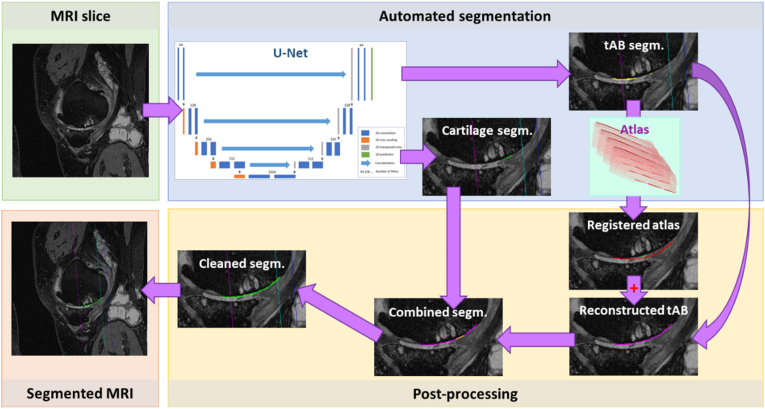
Objective: Denuded areas of subchondral bone (dAB) pose a challenge for fully automated segmentation of articular cartilage and subchondral bone in knees with severe radiographic osteoarthritis using convolutional neural networks (CNNs). Here we propose an automated post-processing relying on a selection-based multi-atlas registration for reconstructing the total area of subchondral bone (tAB) to overcome this issue. We evaluate the agreement, accuracy and longitudinal sensitivity to cartilage change of this novel methodology.
Design: CNN-based models were trained using manual cartilage segmentations from sagittal DESS and coronal FLASH MRI of knees with radiographic (KLG2-4) or severe radiographic osteoarthritis (KLG4 only). These were then applied to KLG4 test knees with manual cartilage segmentations. Automated post-processing was applied to reconstruct missing parts of the tAB and to refine the segmentations, particularly for dABs. The agreement and accuracy of automated cartilage analysis were evaluated using Dice Similarity Coefficients (DSC) and Bland-Altman analyses; sensitivity to one-year change was assessed using the standardized response mean (SRM).
Results: Stronger agreement (DSC 0.80 ± 0.07 to 0.89 ± 0.05) and lower systematic offsets for cartilage thickness (1.2 %-8.4 %) and tAB area (-0.4 %-4.3 %) were observed for CNNs trained on KLG2-4 rather than KLG4 knees; overall, results were superior to those without registration-based post-processing. Sensitivity to change was greatest for manual segmentation of DESS (SRM ≥ -0.69; automated: ≥-0.56) and for automated segmentation of FLASH (≥-0.74; manual ≥-0.44).
Conclusion: CNN-based segmentation combined with registration-based post-processing for accurate delineation of tABs/dABs substantially improves fully-automated (longitudinal) analysis of cartilage and subchondral bone morphology in knees with severe radiographic osteoarthritis.
Keywords: Convolutional neural network; Fully-automated analysis; Imaging; Osteoarthritis; Severe radiographic OA.
© 2025 The Authors.
Conflict of interest statement
Felix Eckstein is co-owner and CEO of Chondrometrics GmbH, a company providing MR image analysis services to academic researchers and to the pharmaceutical industry. He has provided consulting services to Merck KGaA Galapagos/Servier, Kolon Tissuegene, Novartis, Peptinov, Formation Bio, 4P Pharma, Sanofi, and Artialis. Wolfgang Wirth has a part time employment with Chondrometrics GmbH and is a co-owner of Chondrometrics GmbH.
Figures


Similar articles
-
Comparison between coronal FLASH and sagittal double echo steady state MRI in detecting longitudinal cartilage thickness change by fully automated segmentation - Data from the FNIH biomarker cohort.Osteoarthr Cartil Open. 2025 Aug 5;7(3):100657. doi: 10.1016/j.ocarto.2025.100657. eCollection 2025 Sep. Osteoarthr Cartil Open. 2025. PMID: 40822965 Free PMC article.
-
Automated quantitative analysis of peri-articular bone microarchitecture in HR-pQCT knee images.Comput Methods Programs Biomed. 2025 Sep;269:108882. doi: 10.1016/j.cmpb.2025.108882. Epub 2025 Jun 13. Comput Methods Programs Biomed. 2025. PMID: 40532366
-
Evaluation of an automated laminar cartilage T2 relaxation time analysis method in an early osteoarthritis model.Skeletal Radiol. 2025 Mar;54(3):571-584. doi: 10.1007/s00256-024-04786-1. Epub 2024 Sep 4. Skeletal Radiol. 2025. PMID: 39230576 Free PMC article.
-
Mobile bearing vs fixed bearing prostheses for posterior cruciate retaining total knee arthroplasty for postoperative functional status in patients with osteoarthritis and rheumatoid arthritis.Cochrane Database Syst Rev. 2015 Feb 4;2015(2):CD003130. doi: 10.1002/14651858.CD003130.pub3. Cochrane Database Syst Rev. 2015. PMID: 25650566 Free PMC article.
-
Automated devices for identifying peripheral arterial disease in people with leg ulceration: an evidence synthesis and cost-effectiveness analysis.Health Technol Assess. 2024 Aug;28(37):1-158. doi: 10.3310/TWCG3912. Health Technol Assess. 2024. PMID: 39186036 Free PMC article.
Cited by
-
Comparison between coronal FLASH and sagittal double echo steady state MRI in detecting longitudinal cartilage thickness change by fully automated segmentation - Data from the FNIH biomarker cohort.Osteoarthr Cartil Open. 2025 Aug 5;7(3):100657. doi: 10.1016/j.ocarto.2025.100657. eCollection 2025 Sep. Osteoarthr Cartil Open. 2025. PMID: 40822965 Free PMC article.
References
-
- Schnitzer T., Pueyo M., Deckx H., Aar E van der, Bernard K., Hatch S., et al. Evaluation of S201086/GLPG1972, an ADAMTS-5 inhibitor, for the treatment of knee osteoarthritis in ROCCELLA: a phase 2 randomized clinical trial. Osteoarthr. Cartil. 2023;31(7):985–994. - PubMed
-
- Lohmander L.S., Hellot S., Dreher D., Krantz E.F.W., Kruger D.S., Guermazi A., et al. Intraarticular sprifermin (recombinant human fibroblast growth factor 18) in knee osteoarthritis: a randomized, double-blind, placebo-controlled trial. Arthritis Rheumatol. (Hoboken, NJ) 2013;66(7):1820–1831. - PubMed
LinkOut - more resources
Full Text Sources
