The brain on expert medical performance: a systematic review and activation likelihood estimation functional magentic resonance imaging meta-analysis
- PMID: 40697627
- PMCID: PMC12280874
- DOI: 10.1093/psyrad/kkaf019
The brain on expert medical performance: a systematic review and activation likelihood estimation functional magentic resonance imaging meta-analysis
Abstract
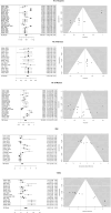
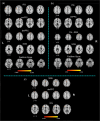
Healthcare systems require the efficient development of expert performance. Several studies have explored the cognitive foundations of medical expert performance, especially in radiology. Studying at the brain level could provide further insight into specific mechanisms mediating medical expert performance. Researchers have recently begun to systematically employ neuroimaging in this field. Most studies focus on specific specializations rather than identifying shared neural substrates across disciplines. This systematic review and activation likelihood estimation (ALE) meta-analysis followed the PRISMA (Preferred Reporting Items for Systematic reviews and Meta-Analyses) guidelines. A total of 297 studies examining neural correlates were identified by comparing expert and novice medical performance. After screening, 22 studies were included in the final analysis. For studies reporting three-dimensional coordinates, ALE meta-analysis revealed consistent involvement of the medial frontal lobe, including the superior frontal gyrus, dorsomedial and ventromedial prefrontal cortex, and inferior frontal and fusiform gyri. Radiology-specific analyses highlighted activation in the ventromedial prefrontal cortex, the left pre-supplementary motor area (pre-SMA), along with the fusiform and opercular inferior frontal gyri. Internal medicine-based studies highlighted involvement of the SMA, inferior frontal gyrus, and dorsomedial prefrontal cortex. Our results revealed involvement, at different levels, of the medial frontal cortex, including the SMA and superior and inferior frontal gyri, which is part of the network relevant for inhibitory control and decision-making. The development of decision-making during the diagnostic process is relevant for the training of future professionals.
Keywords: Medical expert performance; SMA; fMRI; learning; radiology.
© The Author(s) 2025. Published by Oxford University Press on behalf of West China School of Medicine/West China Hospital (WCSM/WCH) of Sichuan University.
Conflict of interest statement
None declared.
Figures


Similar articles
-
Systemic pharmacological treatments for chronic plaque psoriasis: a network meta-analysis.Cochrane Database Syst Rev. 2021 Apr 19;4(4):CD011535. doi: 10.1002/14651858.CD011535.pub4. Cochrane Database Syst Rev. 2021. Update in: Cochrane Database Syst Rev. 2022 May 23;5:CD011535. doi: 10.1002/14651858.CD011535.pub5. PMID: 33871055 Free PMC article. Updated.
-
Sexual Harassment and Prevention Training.2024 Mar 29. In: StatPearls [Internet]. Treasure Island (FL): StatPearls Publishing; 2025 Jan–. 2024 Mar 29. In: StatPearls [Internet]. Treasure Island (FL): StatPearls Publishing; 2025 Jan–. PMID: 36508513 Free Books & Documents.
-
The Role of the Amygdala in Facial Trustworthiness Processing: A Systematic Review and Meta-Analyses of fMRI Studies.PLoS One. 2016 Nov 29;11(11):e0167276. doi: 10.1371/journal.pone.0167276. eCollection 2016. PLoS One. 2016. PMID: 27898705 Free PMC article.
-
Short-Term Memory Impairment.2024 Jun 8. In: StatPearls [Internet]. Treasure Island (FL): StatPearls Publishing; 2025 Jan–. 2024 Jun 8. In: StatPearls [Internet]. Treasure Island (FL): StatPearls Publishing; 2025 Jan–. PMID: 31424720 Free Books & Documents.
-
Non-pharmacological interventions for improving language and communication in people with primary progressive aphasia.Cochrane Database Syst Rev. 2024 May 29;5(5):CD015067. doi: 10.1002/14651858.CD015067.pub2. Cochrane Database Syst Rev. 2024. PMID: 38808659 Free PMC article.
References
-
- Akyürek EG, Kappelmann N, Volkert M et al. (2017) What you see is what you remember: visual chunking by temporal integration enhances working memory. J Cogn Neurosci. 29:2025–36. - PubMed
-
- Amunts K, Schlaug G, Jäncke L et al. (1997) Motor cortex and hand motor skills: structural compliance in the human brain. Hum Brain Mapp. 5:206–15. - PubMed
Publication types
LinkOut - more resources
Full Text Sources
