Review
. 2025 Jul 22:29:0219.
doi: 10.34133/bmr.0219. eCollection 2025.

Metal-Based Regenerative Strategies for Peripheral Nerve Injuries: From Biodegradable Ion Source to Stable Conductive Implants

Affiliations
Review

Metal-Based Regenerative Strategies for Peripheral Nerve Injuries: From Biodegradable Ion Source to Stable Conductive Implants

Hyewon Kim et al. Biomater Res. .

Abstract

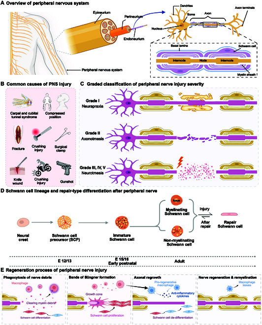
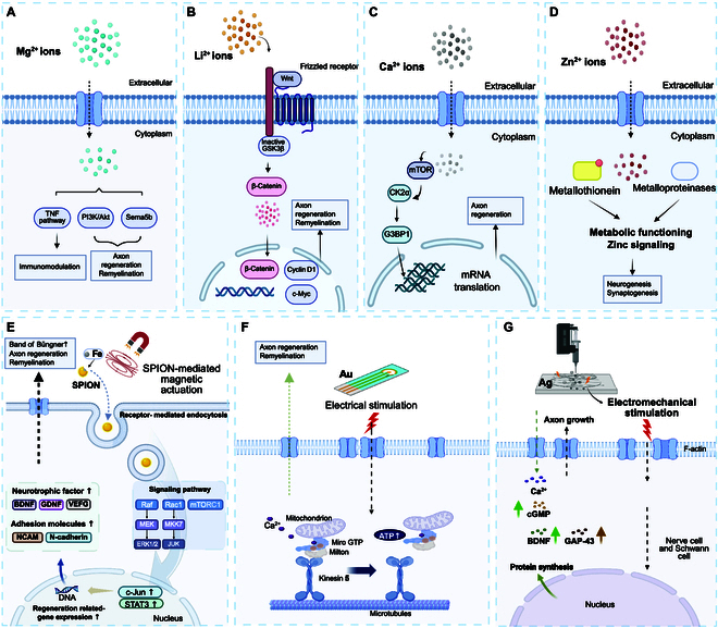
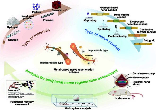
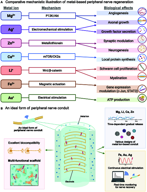
Peripheral nerve injury is a common health issue in modern aging societies, with the only treatment available being autograft transplantation. Unfortunately, autograft is often limited due to donor availability and immune rejection. Additionally, the peripheral nervous system has limited regenerative capacity, making the treatment of peripheral nerve injuries challenging. Metal-based regenerative medicine and tissue engineering strategies provide advanced solutions to the problem. Metal-based biomaterials such as conduits, filaments, alloys, hydrogels, and ceramics can deliver biofunctional metal ions and promote axonal growth and functional recovery. In parallel, metal-based electromagnetic stimulation demonstrates potential for nerve regeneration and inflammation regulation. The potential of metal-based biomaterials in promoting peripheral nerve regeneration highlights the need for further research in tissue engineering and regenerative medicine. However, rapid degradation, long-term biocompatibility, and necessary optimization regarding injury types remain to be explored. This review summarizes the reported metal-based biomaterials utilized in peripheral nerve regeneration research. The aim is to showcase advanced technologies available in the field, which may potentially become a viable alternative to autografts, offering transformative applications in the regenerative medical field.


Conflict of interest statement

Competing interests: The authors declare that they have no competing interests.

Figures

Fig. 1.
Fig. 1.
Structural organization, classification, cellular dynamics, and regeneration process of PNI. (A) Schematic representation of the peripheral nervous system anatomy, highlighting the hierarchical organization of connective tissue layers, which are epineurium, perineurium, and endoneurium surrounding individual nerve fascicles. The microarchitecture includes axons ensheathed by Schwann cells and organized into myelinated internodal segments. (B) Common etiologies of PNIs include mechanical compression (e.g., carpal or cubital tunnel syndrome), blunt trauma (e.g., fractures and crush injuries), and penetrating injuries (e.g., surgical clamps, gunshot wounds, and knife lacerations). (C) Graded classification of PNI severity based on Sunderland’s scale, ranging from grade I (neuropraxia: transient conduction block without axonal damage), grade II (axonotmesis: axonal disruption with preserved connective tissue architecture), to grades III to V (neurotmesis: progressive structural damage involving endoneurial, perineurial, and epineurial disruption). (D) Developmental lineage and injury-induced plasticity of Schwann cells. Schwann cells arise from neural crest cells via a precursor stage and differentiate into myelinating or nonmyelinating phenotypes. Upon injury, mature Schwann cells dedifferentiate into a repair phenotype, enabling axonal guidance, debris clearance, and trophic support, followed by redifferentiation during regeneration. (E) Sequential events during peripheral nerve regeneration, including macrophage-mediated debris clearance, Schwann cell reprogramming and proliferation, formation of bands of Büngner, axonal elongation, and eventual remyelination, culminating in functional restoration of nerve integrity.
Fig. 2.
Fig. 2.
Illustration of molecular mechanisms of metal-based peripheral nerve regeneration. (A) Mg2+ enters cells via transporters, activating key pathways such as TNF and PI3K/Akt, and stimulating Sema5b to support immunomodulation, axon regeneration, and remyelination. (B) Li+ activates the Wnt/β-catenin signaling pathway, where the frizzled receptor inactivates GSK3β, enabling β-catenin to up-regulate c-Myc and cyclin D1 for facilitating axon regeneration and remyelination. (C) Role of calcium ions (Ca2+) in axon regeneration through mTOR pathway. (D) Role of zinc ions (Zn2+) in synaptic transmission and neurogenesis. Illustration of iron and gold ion-based promotes nerve regeneration. (E) Schematic illustration of the mechanism by which SPION-mediated magnetic actuation promotes peripheral nerve regeneration. SPIONs exert mechanical stimulation inside the cell, which activates intracellular signaling pathways promoting the formation of bands of Büngner, axon regeneration, and remyelination, which are critical for peripheral nerve repair. (F) Gold (Au)-based electrodes provide electrical stimulation, enhancing axon growth by boosting mitochondrial transport via Kinesin 5, Miro GTP, and Milton, which increases ATP production to support neuronal repair [64]. (G) Ag-based electrodes, when subjected to electrical stimulation, up-regulate the secretion of growth factors such as FGF, CNTF, and GDNF, while electromechanical stimulation enhances axon growth and cell proliferation.
Fig. 3.
Fig. 3.
Overview of metal-based strategies and evaluation methods for peripheral nerve regeneration. The schematic illustrates an integrated perspective on material types, nerve conduit design, and assessment techniques used in metal-based peripheral nerve regeneration research. At the center, metal-based nerve regeneration mechanisms are categorized into biodegradable and implantable systems. Biodegradable metals (e.g., Mg2+, Zn2+, Ca2+, and Li+) release bioactive ions that influence regeneration pathways. Implantable metals (e.g., Au, Ag, and Fe) may interact with electric or magnetic fields to stimulate axonal growth and inflammation modulation. At the top left (brown), biomaterials are classified by physical state and hardness, ranging from soft solutions and hydrogels to ion-doped polymers, particles, filaments, and ceramics. At the top right (blue) are various fabrication methods and types of NGCs, including hydrogel-based conduits, metal-coated scaffolds, electrospun nanofibers, conductive polymer conduits, and autografts. Fabrication techniques like 3D printing, sputtering, and electrospinning enable controlled architecture and functional integration. Finally, at the bottom, an in vivo experimental setup showing nerve conduit implantation bridging proximal and distal nerve stumps in a rodent sciatic nerve injury model. Methods for functional recovery assessment, including walking track analysis and calculation of the SFI based on hindlimb footprint parameters and sciatic nerve distribution.
Fig. 4.
Fig. 4.
(A) Comparative mechanistic illustration of metal-based peripheral nerve regeneration and an ideal form of peripheral nerve conduit. The relationship between representative metal ions, their associated molecular mechanisms, and the resulting biological effects in the context of peripheral nerve regeneration. The visual layout enables cross-comparison of how different ions contribute to key processes such as axonal growth, Schwann cell proliferation, and myelination through distinct mechanistic pathways. (B) Illustration of an ideal metal-based nerve conduit. An ideal peripheral nerve conduit should be excellent biocompatible, capable of releasing metal ions gradually, and long-term conductive to stimulate nerve cells when necessary.

Similar articles

  • Bioengineered nerve conduits and wraps for peripheral nerve repair of the upper limb.
    Thomson SE, Ng NY, Riehle MO, Kingham PJ, Dahlin LB, Wiberg M, Hart AM. Thomson SE, et al. Cochrane Database Syst Rev. 2022 Dec 7;12(12):CD012574. doi: 10.1002/14651858.CD012574.pub2. Cochrane Database Syst Rev. 2022. PMID: 36477774 Free PMC article.
  • Management of urinary stones by experts in stone disease (ESD 2025).
    Papatsoris A, Geavlete B, Radavoi GD, Alameedee M, Almusafer M, Ather MH, Budia A, Cumpanas AA, Kiremi MC, Dellis A, Elhowairis M, Galán-Llopis JA, Geavlete P, Guimerà Garcia J, Isern B, Jinga V, Lopez JM, Mainez JA, Mitsogiannis I, Mora Christian J, Moussa M, Multescu R, Oguz Acar Y, Petkova K, Piñero A, Popov E, Ramos Cebrian M, Rascu S, Siener R, Sountoulides P, Stamatelou K, Syed J, Trinchieri A. Papatsoris A, et al. Arch Ital Urol Androl. 2025 Jun 30;97(2):14085. doi: 10.4081/aiua.2025.14085. Epub 2025 Jun 30. Arch Ital Urol Androl. 2025. PMID: 40583613 Review.
  • Short-Term Memory Impairment.
    Cascella M, Al Khalili Y. Cascella M, et al. 2024 Jun 8. In: StatPearls [Internet]. Treasure Island (FL): StatPearls Publishing; 2025 Jan–. 2024 Jun 8. In: StatPearls [Internet]. Treasure Island (FL): StatPearls Publishing; 2025 Jan–. PMID: 31424720 Free Books & Documents.
  • Sexual Harassment and Prevention Training.
    Cedeno R, Bohlen J. Cedeno R, et al. 2024 Mar 29. In: StatPearls [Internet]. Treasure Island (FL): StatPearls Publishing; 2025 Jan–. 2024 Mar 29. In: StatPearls [Internet]. Treasure Island (FL): StatPearls Publishing; 2025 Jan–. PMID: 36508513 Free Books & Documents.
  • Scaffold design considerations for peripheral nerve regeneration.
    Yu L, Bennett CJ, Lin CH, Yan S, Yang J. Yu L, et al. J Neural Eng. 2024 Jul 23;21(4):10.1088/1741-2552/ad628d. doi: 10.1088/1741-2552/ad628d. J Neural Eng. 2024. PMID: 38996412 Free PMC article. Review.

References

    1. Widodo W, Aprilya D, Satria O. Regenerative medicine: A new horizon in peripheral nerve injury and repair. Orthop Rev. 2025;17:133572. - PMC - PubMed
    1. Magneli M, Axenhus M. Epidemiology and regional variance of traumatic peripheral nerve injuries in Sweden: A 15-year observational study. PLOS ONE. 2024;19(10): Article e0310988. - PMC - PubMed
    1. Kim SJ, Kwon YM, Ahn SM, Lee JH, Lee C-H. Epidemiology of upper extremity peripheral nerve injury in South Korea, 2008 to 2018. Medicine. 2022;101(48): Article e31655. - PMC - PubMed
    1. Murphy RNA, Schoulepnikoff C, Chen JHC, Columb MO, Bedford J, Wong JK, Reid AJ. The incidence and management of peripheral nerve injury in England (2005–2020). J Plast Reconstr Aesthet Surg. 2023;80:75–85. - PubMed
    1. Hussain G, Wang J, Rasul A, Anwar H, Qasim M, Zafar S, Aziz N, Razzaq A, Hussain R, Aguilar J-LG, et al. Current status of therapeutic approaches against peripheral nerve injuries: A detailed story from injury to recovery. Int J Biol Sci. 2020;16(1):116. - PMC - PubMed

LinkOut - more resources