. 2025 Apr 29;6(1):374-390.
doi: 10.1089/neur.2024.0168. eCollection 2025.

Chronic Neurobehavioral and Neuropathological Consequences of Repeated Blast Exposure in P301S Transgenic Tau Rats

Affiliations

Chronic Neurobehavioral and Neuropathological Consequences of Repeated Blast Exposure in P301S Transgenic Tau Rats

Claire Robey et al. Neurotrauma Rep. .

Abstract

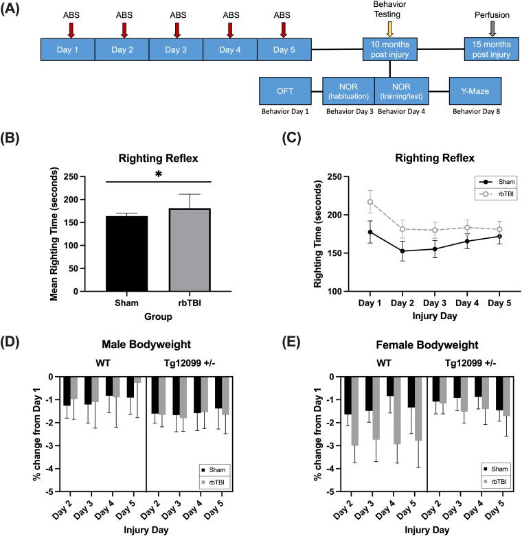
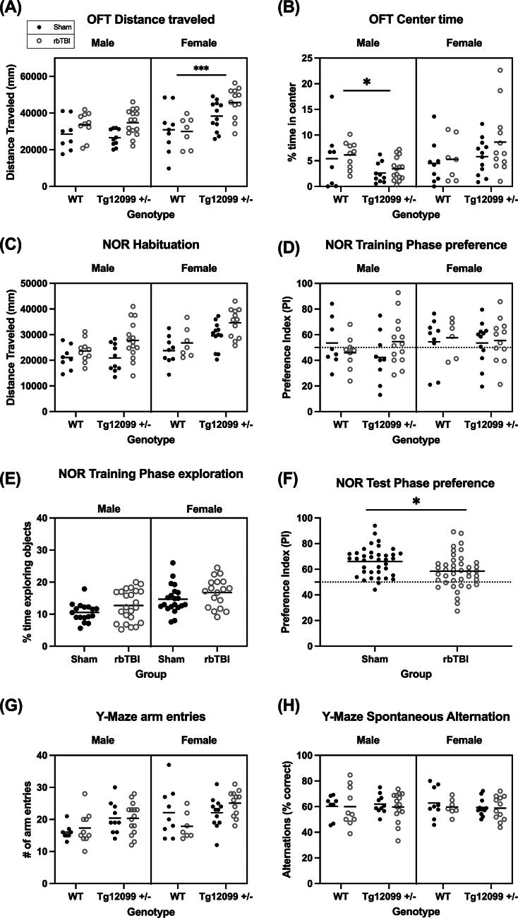
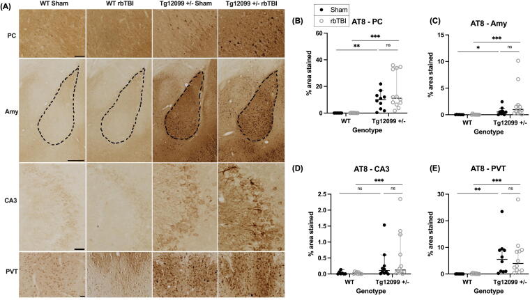
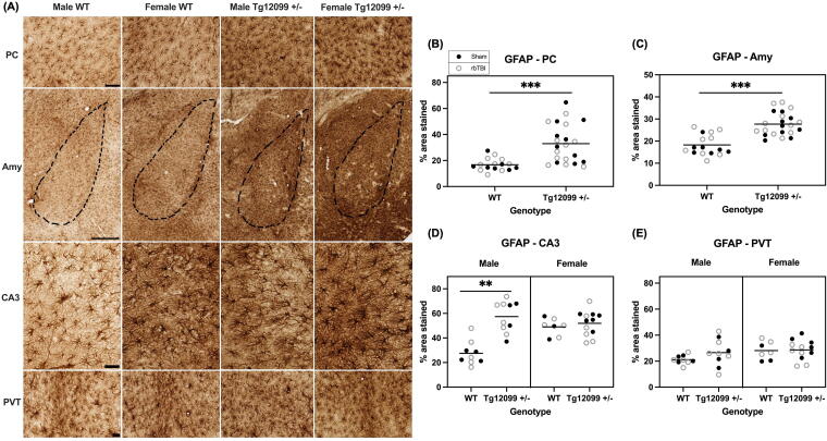
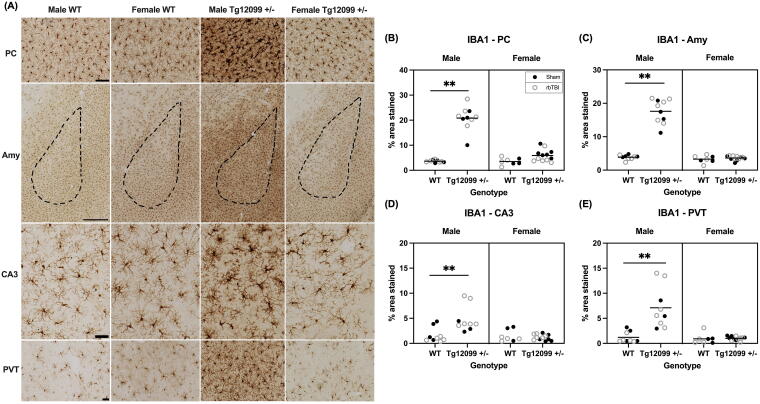
Repeated blast traumatic brain injury (rbTBI) is linked to dementia risk, potentially due to abnormal tau accumulation, although a definitive causal relationship remains elusive. This study aims to develop a model of rbTBI-induced tauopathy. We utilized wild-type (WT) rats and rats that are heterozygous for the mutated P301S human tau gene (Tg12099 +/-), the presence of which increases the propensity to develop tau neuropathology. At 2-3 months of age, rats were exposed to five blasts using the Advanced Blast Simulator or sham procedures. Behavioral and histological outcomes were evaluated at 10 and 15 months post-injury, respectively. The open field test revealed increased activity in blast-injured animals compared with sham. Tg12099 +/- females exhibited greater travel distances than WT females, while male activity levels did not differ by genotype. The novel object recognition test indicated impaired recognition memory in blast-injured animals, which was unrelated to genotype. There was a greater accumulation of phosphorylated tau in several brain regions of Tg12099 +/- rats compared with WT rats, yet no observable blast injury effect. Blast did not alter astro- and microgliosis, but increased astrogliosis was observed in Tg12099 +/- rats compared with WT rats in a region-dependent manner. We observed sex-dependent changes in microgliosis within the Tg12099 +/- group, with male Tg12099 +/- rats exhibiting increased IBA1 immunostaining compared with females. No such sex difference was observed in WT rats. Our findings suggest that while rbTBI can induce persistent behavioral deficits in rats, it does not exacerbate neuropathology in Tg12099 rats.

Keywords: aging; behavior; neuropathology; sex differences; tau; traumatic brain injury.


Figures

FIG. 1.
FIG. 1.
Experimental timeline, righting time, and body weight. (A) Schematic of the experimental procedure timeline. (B) Mean righting time was longer in blast-injured rats compared with sham rats (p = 0.032). (C) There was no effect of Day on righting time (p = 0.177). (D–E) Percent change in body weight from Day 1 (preblast) in male (D) and female (E) rats. All groups lost minimal weight from Day 1, but changes did not differ based on injury, genotype, or sex. n = 7–16 rats per group. *indicates p < 0.05. Data depict mean and SEM. ABS, Advanced Blast Simulator; rbTBI, repeated blast traumatic brain injury; SEM, standard error of the mean; Tg12099 +/−, transgenic heterozygous P301S Tau rat; WT, wild-type.
FIG. 2.
FIG. 2.
Behavior testing at 10 months post-injury. (A) OFT distance traveled. Blast-injured animals traveled a greater distance compared with sham animals. There was also a main effect of sex (F > M) and genotype (Tg12099 +/− > WT). In addition, there was a significant interaction between genotype × sex. Tg12099 +/− females traveled farther than WT females, while activity levels did not differ by genotype in males. (B) OFT center time. There was a significant interaction between genotype and sex. Tg12099 +/− males spent less time in the center than WT males, while center time did not differ by genotype in females. (C) NOR habituation. There were main effects of injury, sex, and genotype where activity levels were increased in blast-injured animals compared with sham, females traveled greater distances than males, and Tg12099 +/− rats traveled greater distances than WT rats. (D) NOR training phase preference. No differences were seen in object preference when exploring two identical objects during the training phase. The dashed line depicts 50% PI indicating no preference. (E) NOR training phase exploration. Injured animals spent more time exploring the identical objects compared with sham (p = 0.0127) and females spent more time exploring the identical objects than males (p = 0.0001). (F) NOR test phase. Injured animals had a lower PI than sham animals, indicating less preference for the novel object (p = 0.0108). (G) Y-maze arm entries. Females had a greater number of arm entries compared with males (p = 0.0015), and Tg12099 +/− rats had a greater number of arm entries compared with WT rats (p = 0.0042). (H) Y-maze spontaneous alternation. No significant differences were observed for % correct alternation. Lines depict means (n = 7–16 rats per group). *p < 0.05, ***p < 0.001. Data were combined for presentation purposes in (E) (no genotype effect) and (F) (no genotype or sex effect). NOR, novel object recognition; OFT, open field test; PI, preference index; rbTBI, repeated blast traumatic brain injury.
FIG. 3.
FIG. 3.
Immunohistochemistry for AT8 (phosphorylated tau) at 15 months post-injury (17–19 months old). (A) Representative images of piriform cortex (PC), amygdala (Amy), cornu ammonis 3 region of the hippocampus (CA3), and paraventricular nucleus of the thalamus (PVT). Scale bars = 100 µm for PC, CA3, and PVT; scale bar = 500 µm for Amy. (B–E) Quantification of percentage of area stained in the PC, Amy, CA3, and PVT. Both injured and sham Tg12099 +/− rats exhibited greater AT8 staining compared with their respective WT controls in the PC, Amy, and PVT. In CA3, only injured Tg12099 +/− rats exhibited greater AT8 staining compared with injured WT rats (D). The Kruskal—Wallis rank test, followed by Dunn’s multiple comparison test, was used to analyze each region. There were no sex differences so male and female data were pooled for analysis. Lines represent median and error bars indicate 95% CI (n = 7–12 rats per group); ns = not significant, *p < 0.05, **p < 0.01, ***p < 0.001. AT8, phosphorylated tau (Serine 202/Threonine 205).
FIG. 4.
FIG. 4.
Immunohistochemistry for GFAP at 15 months post-injury (17–19 months old). (A) There were no injury differences in any brain region so representative images depict both sex and genotype in the piriform cortex (PC), amygdala (Amy), cornu ammonis 3 region of the hippocampus (CA3), and paraventricular nucleus of the thalamus (PVT). Scale bars = 100 µm for PC, CA3, and PVT; scale bar = 500 µm for Amy. (B–D) Quantification of percentage of area stained in the PC, Amy, CA3, and PVT. Tg12099 +/− rats exhibited higher GFAP-positive area stained compared with WT rats in the PC and Amy. In CA3, male Tg12099 +/− rats had higher GFAP than male WT rats, but in female rats both WT and Tg12099 +/− GFAP levels were higher than male WT. (D) No differences were seen in astrogliosis in the PVT. A three-way ANOVA was used to analyze each region. Lines indicate mean (n = 7–12 rats per group). Data were combined for presentation purposes in (B) and (C) (no injury or sex effect), (D) and (E) (no injury effect). GFAP, glial fibrillary acidic protein.
FIG. 5.
FIG. 5.
Immunohistochemistry for IBA1 at 15 months post-injury (17–19 months old). (A) Representative images by sex and genotype in the piriform cortex (PC), amygdala (Amy), CA3 region of the hippocampus, and paraventricular nucleus of the thalamus (PVT). Scale bars = 100 µm for PC, CA3, and PVT; scale bar = 500 µm for Amy. (B–E) Quantification of percentage area stained in the PC, Amy, CA3, and PVT. Male Tg12099 +/− rats exhibited higher IBA1-positive area stained compared with male WT rats and both female groups in all four brain regions. In the CA3 region, there was also an injury and genotype interaction, where injured Tg12099 +/− rats had higher levels of IBA1 compared with injured WT rats, but there was no genotype difference in sham rats (D). A three-way ANOVA was used to analyze each region. Lines indicate mean (n = 3–12 rats per group). Data were combined for presentation purposes in (no injury effect). IBA1, ionized calcium-binding adaptor molecule 1.

Similar articles

References

    1. Okie S. Traumatic brain injury in the war zone. N Engl J Med 2005;352(20):2043–2047; doi: 10.1056/NEJMp058102 - DOI - PubMed
    1. Kontos AP, Kotwal RS, Elbin R, et al. Residual effects of combat-related mild traumatic brain injury. J Neurotrauma 2013;30(8):680–686; doi: 10.1089/neu.2012.2506 - DOI - PubMed
    1. McKee AC, Robinson ME. Military‐related traumatic brain injury and neurodegeneration. Alzheimers Dement 2014;10(3 Suppl):S242–S253; doi: 10.1016/j.jalz.2014.04.003 - DOI - PMC - PubMed
    1. Phipps H, Mondello S, Wilson A, et al. Characteristics and impact of US military blast-related mild traumatic brain injury: A systematic review. Front Neurol 2020;11:559318. - PMC - PubMed
    1. Brenner LA, Ivins BJ, Schwab K, et al. Traumatic brain injury, posttraumatic stress disorder, and postconcussive symptom reporting among troops returning from Iraq. J Head Trauma Rehabil 2010;25(5):307–312. - PubMed

LinkOut - more resources