

Ganglion Cell Layer Thickness as a Biomarker for Amyotrophic Lateral Sclerosis Functional Outcome: An OCT study
- PMID: 40698100
- PMCID: PMC12277982
- DOI: 10.22336/rjo.2025.32
Ganglion Cell Layer Thickness as a Biomarker for Amyotrophic Lateral Sclerosis Functional Outcome: An OCT study
Abstract
Aim: This study aims to evaluate various optical coherence tomography (OCT) parameters in patients diagnosed with amyotrophic lateral sclerosis (ALS).
Methods: Assessment of BCVA was done using Snellen charts, and subjective refraction was done to achieve a BCVA for distance and near. Measurement of intraocular pressure (IOP) was done with Goldman applanation tonometry. Stereoscopic fundus examination was performed using a 90D lens to assess the status of the optic nerve and retina, ruling out any ocular pathology. The patients were then subjected to OCT scanning to measure optic nerve head and macular parameters. Optical coherence tomography was performed using CIRRUS™ HD OCT (500-21822) (version 8.0.0.518) (Carl Zeiss Meditec, Dublin, CA, USA). The analyzed area was centered manually, and the absence of segmentation errors was confirmed for each scan.
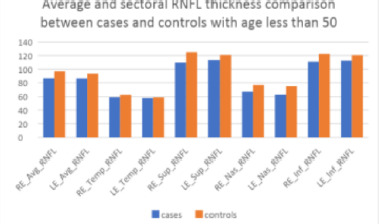
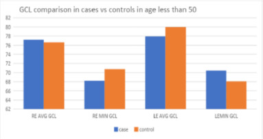
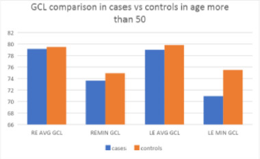
Results: RE Avg RNFL and LE Avg RNFL showed weak correlations with ALSFRS, indicated by Pearson Correlation coefficients of 0.073 and -0.026, respectively. The p-values (0.637 and 0.86) suggested that these correlations were not statistically significant. RE Avg GCL and LE Avg GCL, on the other hand, exhibited moderate positive correlations with ALSFRS scores, with correlation coefficients of 0.337 (RE) and 0.389 (LE). These correlations were statistically significant, as indicated by p-values of 0.021 and 0.006, respectively, suggesting a substantial association between GCL thickness and ALS functional outcomes.
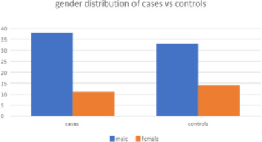
Discussion: All patients in our study were clinically diagnosed cases of ALS, as per the El Escorial criteria. Age group-wise analysis showed statistically significant thinning overall as well as quadrant-wise RNFL parameters in patients less than 50 years compared to age-matched controls, indicating that the pathological process occurring in larger motor neurons in ALS might also be happening in smaller sensory neurons of the retina, causing thinning, which was not due to age-related process. Although GCIPL thinning was occurring in our cases, though statistically not significant compared to control, the significant positive correlation observed between GCIPL and ALS functional outcome and between RNFL and GCIPL measurements highlighted the fact that though the axonal degeneration in retinal neurons might not be translating to the same extent in ganglion cells in ALS, the subtle thinning of GCIPL correlated strongly with functional disability in patients with ALS, implying better functional scores with higher values of GCIPL parameters.
Conclusion: In summary, GCL measurements in both eyes showed a notable relationship with ALSFRS, whereas RNFL did not appear to correlate significantly.
Keywords: AD = Alzheimer's disease; ALS; ALS = amyotrophic lateral sclerosis; ALSFRS; ALSFRS = ALS functional rating scale; BCVA = best corrected visual acuity; CMT = central macular thickness; CNS = central nervous system; CST = central subfield thickness; DTI = diffusion tensor imaging; EEC = El Escorial criteria; ELM OLM = external or outer limiting membrane; EMG = electromyography; ESA = existing segmentation algorithm; ETDRS = early treatment for diabetic retinopathy study; FD OCT = Fourier domain optical coherence tomography; FEV 1 = forced expiratory volume at 1 sec; FVC = forced vital capacity; FWHM = full width half maximum; GCIPL; GCIPL = ganglion cell inner plexiform layer; HC = healthy controls; HCDR = horizontal cup to disc ratio; ILM = internal limiting membrane; IMR = inner macular ring; INL = inner nuclear layer; IOP = intraocular pressure; IPL = inner plexiform layer; IS OS = inner segment outer segment junction; LMN = lower motor neuron; MND = motor neuron disease; MRI = magnetic resonance imaging; MT = macular thickness; NCV = nerve conduction velocity; NDD = neurodegenerative disorder; NFL = nerve fibre layer; OCT = optical coherence tomography; OMR = outer macular ring; ONH = optic nerve head; ONL = outer nuclear layer; OPL = outer plexiform layer; OPTN = optineurin; PD = Parkinson’s disease; PFN = profilin gene; RNFL; RNFL = retinal nerve fibre layer; RPE = retinal pigment epithelium; SD OCT = spectral domain optical coherence tomography; SLD = super luminescent diode; SNR = signal to noise ratio; SPSS = statistical package for social sciences; SS OCT = swept source optical coherence tomography; TBK 1 = TANK binding kinase 1 gene; TD OCT = time domain optical coherence tomography; UCVA = uncorrected visual acuity; UMN = upper motor neuron; VCDR = vertical cup to disc ratio; VEP = visually evoked potential; functional outcome; pRNFL = peripapillary RNFL; structural outcome.
© 2025 The Authors.
Conflict of interest statement
The authors state no conflict of interest.
Figures
Similar articles
-
Regional retinal vulnerability in multiple sclerosis: integrating OCT, MRI, and clinical data for enhanced diagnosis and automated monitoring.Rom J Morphol Embryol. 2025 Jan-Mar;66(1):119-130. doi: 10.47162/RJME.66.1.11. Rom J Morphol Embryol. 2025. PMID: 40384198 Free PMC article.
-
Optic nerve head and fibre layer imaging for diagnosing glaucoma.Cochrane Database Syst Rev. 2015 Nov 30;2015(11):CD008803. doi: 10.1002/14651858.CD008803.pub2. Cochrane Database Syst Rev. 2015. PMID: 26618332 Free PMC article.
-
Retinal layer segmentation in multiple sclerosis: a systematic review and meta-analysis.Lancet Neurol. 2017 Oct;16(10):797-812. doi: 10.1016/S1474-4422(17)30278-8. Epub 2017 Sep 12. Lancet Neurol. 2017. PMID: 28920886
-
Macular versus Retinal Nerve Fiber Layer Parameters for Diagnosing Manifest Glaucoma: A Systematic Review of Diagnostic Accuracy Studies.Ophthalmology. 2016 May;123(5):939-49. doi: 10.1016/j.ophtha.2015.12.041. Epub 2016 Feb 15. Ophthalmology. 2016. PMID: 26891880
-
Optical coherence tomography (OCT) for detection of macular oedema in patients with diabetic retinopathy.Cochrane Database Syst Rev. 2015 Jan 7;1(1):CD008081. doi: 10.1002/14651858.CD008081.pub3. Cochrane Database Syst Rev. 2015. PMID: 25564068 Free PMC article.
References
-
- Soto C, Estrada LD. Protein misfolding and neurodegeneration. Arch. Neurol. 2008;65:184–189. - PubMed
-
- Blanks JC, Schmidt SY, Torigoe Y, Porrello KV, Hinton DR, Blanks RH. Retinal pathology in Alzheimer’s disease: Regional neuron loss and glial changes in GCL. Neurobiol. Aging. 1996;17:385–395. - PubMed
-
- Blanks JC, Torigoe Y, Hinton DR, Blanks RH. Retinal pathology in Alzheimer’s disease. I. Ganglion cell loss in foveal/parafoveal retina. Neurobiol. Aging. 1996;17:377–384. - PubMed
-
- Hinton DR, Sadun AA, Blanks JC, Miller CA. Optic-nerve degeneration in Alzheimer’s disease. N. Engl. J. Med. 1986;315:485–487. - PubMed
MeSH terms
Substances
LinkOut - more resources
Full Text Sources
Medical
Research Materials
Miscellaneous