. 2025 Jul;15(7):3708-3724.
doi: 10.1016/j.apsb.2025.05.003. Epub 2025 May 13.

CMD-OPT model enables the discovery of a potent and selective RIPK2 inhibitor as preclinical candidate for the treatment of acute liver injury

Affiliations

CMD-OPT model enables the discovery of a potent and selective RIPK2 inhibitor as preclinical candidate for the treatment of acute liver injury

Yong Chen et al. Acta Pharm Sin B. 2025 Jul.

Abstract

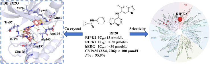
Acute liver injury (ALI) serves as a critical precursor and major etiological factor in the progression and ultimate manifestation of various hepatic disorders. The prevention and treatment of ALI is still a serious global challenge. Given the limited therapeutic options for ALI, exploring novel targeted therapeutic agents becomes imperative. The potential therapeutic efficacy of inhibiting RIPK2 is highlighted, as it may provide significant benefits by attenuating the MAPK pathway and NF-κB signaling. Herein, we propose a CMD-OPT model, a two-stage molecular optimization tool for the rapid discovery of RIPK2 inhibitors with optimal properties. Compound RP20, which targets the ATP binding site, demonstrated excellent kinase specificity, ideal oral pharmacokinetics, and superior therapeutic effects in a model of APAP-induced ALI, positioning RP20 as a promising preclinical candidate. This marks the first application of RIPK2 inhibitors in ALI treatment, opening a novel therapeutic pathway for clinical applications. These results highlight the efficacy of the CMD-OPT model in producing lead compounds from known active molecules, showcasing its significant potential in drug discovery.

Keywords: Acute liver injury; Anti-inflammatory; CMD-OPT; Candidate; Co-crystal; Drug discovery; Kinase specificity; RIPK2 inhibitors.


Conflict of interest statement

The authors declare no conflicts of interest.

Figures

Image 1
Graphical abstract
Figure 1
Figure 1
Representative RIPK2 inhibitors.
Figure 2
Figure 2
Framework and schematic workflow for designing RIPK2 inhibitors using CMD-OPT model.
Scheme 1
Scheme 1
Synthetic approach to RS1-13, 17, 18, 20. (a) p-TSA, IPA, 80 °C; (b) The corresponding borate ester or borate, K2CO3, PdCl2(dppf), Dioxane/EtOH/H2O, 80 °C; (c) TFA, DCM, rt; KOH, H2O; (d) The corresponding anhydride or isocyanate, DCM/MeOH, rt.
Scheme 2
Scheme 2
Synthetic approach to RS14, 15, 16, 19. (a) p-TSA, IPA, 80 °C or NaOH, H2O, 80 °C; (b) 286961-14-6, K2CO3, PdCl2(dppf), Dioxane/EtOH/H2O, 80 °C; (c) TFA, DCM, rt; KOH, H2O.
Scheme 3
Scheme 3
Synthetic approach of intermediate 12. (a) BzCl, CH3CN, 50 °C; (b) Fe, NH4Cl, MeOH/H2O, 65 °C; (c) Br2, KSCN, CH3COOH, rt; (d) tert-Butyl nitrite, THF, 65 °C; (e) 70% H2SO4, 100 °C.
Scheme 4
Scheme 4
Synthetic approach to RP1-29. Reagents and conditions: (a) p-TSA, IPA, 80 °C; (b) The corresponding borate ester or borate, K2CO3, PdCl2 (dppf), Dioxane/EtOH/H2O, 80 °C; (c) TFA, DCM, rt; KOH, H2O; (d) The corresponding anhydride or isocyanate, DCM/MeOH, rt.
Figure 3
Figure 3
Inhibition of RIPK2 activity delays NF-κB activation and RIPK2 ubiquitination. (A) THP-1 cells were either untreated or pre-treated with RP20 and then stimulated with MDP for specified durations. Ubiquitin conjugates were purified using TUBEs, and both lysates and pull-down samples were subjected to WB analysis using the specified antibodies. (B) THP-1 cells were treated with RP20 (2 μmol/L) or left untreated for 0.5 h before stimulation with L18-MDP (200 ng/mL). Cell lysates were harvested at indicated time points and analyzed by WB for p-p65, total p65, p-p38, total p38, p-JNK. (C) BMDMs were treated with RP20 (2 μmol/L) or left untreated for 0.5 h prior to stimulation with L18-MDP (200 ng/mL). Cell lysates were harvested at specified time points and analyzed by WB for p-p65, total p65, p-p38, total p38, p-JNK (n = 3).
Figure 4
Figure 4
Cocrystal structure of RP20 bound within the ATP binding pocket of RIPK2 kinase (PDB: 8X2O). (A) Overview of cocrystal structure of RIPK2 in complex with compound RP20. (B) Detailed depiction of the ATP binding pockets with surrounding key residues depicted as stick models. Hydrogen bonds are indicated by green dashed lines, and the cation–π interaction is denoted by yellow dashed line. (C) Density map of RP20 within the binding site. (D) Protein surface of RIPK2 with the binding structure of RP20. (E) The Pair Interaction Energy (PIE) diagram for RP20 was constructed using the method within a 4.5 Å radius of the residues.
Figure 5
Figure 5
RP20 is a selective inhibitor of RIPK2. (A) The dose–response curves of compound RP20 and GSK2983559 against RIPK2. (B) The kinase selectivity evaluation of compound RP20 at a concentration of 1 μmol/L at Eurofins.
Figure 6
Figure 6
RP20 inhibits NOD2 signaling in vivo and has a beneficial effect on an ALI model. (A) Pharmacodynamic study of RP20 on MDP induced peritonitis model in C57BL/6 mice. 8-week female C57BL/6 mice were sacrificed after 4 h of MDP (100 μg/piece) induction and the serum were obtained. IL-6 levels in serum using ELISA kits. n = 10 (Control, n = 6), mean ± SD. (B) Effects of RP20 and GSK2983559 on APAP-induced ALI model. 6–8 weeks C57BL/6 mice were sacrificed after 12 h of APAP (500 mg/kg) induction and the liver tissues were obtained. H&E staining after fixation with 10% paraformaldehyde. Scale bar, 100 μm. (C) ALT and AST levels by blood biochemical analyzer; n = 6, mean ± SD. (D, E) TNF-α, IL-1β and IL-6 levels in serum using ELISA kits. n = 3, mean ± SD; TNF-α, IL-1β and IL-6 mRNA levels in the liver were detected by QPCR. (F) Statistical chart of liver necrosis area; detection of GSH level in liver tissue using T-GSH kit. n = 3, mean ± SD. Compared with the control group, #P < 0.05, ###P < 0.001; compared with the APAP model group, ∗P < 0.05, ∗∗∗P < 0.001; ns means no significant difference.
Figure 7
Figure 7
RP20 has hepatoprotective effects on ALI models and exhibits liver-targeting properties. (A) TUNEL immunohistochemical staining of liver cells across various experimental groups. Scale bar, 50 μm. (B) Liver cell apoptosis statistics. n = 3, mean ± SD. (C) RP20 inhibits NF-κB and MAPK (p-JNK, p-ERK, p-p65) in liver tissues from treatment groups. (D) Tissue distribution of RP20 in mice with ALI. 6–8 weeks C57BL/6 mice were sacrificed after 2, 4, 8 h after APAP (500 mg/kg) induction and the serum were obtained. n = 5, mean ± SD. (E) ALT and AST levels by blood biochemical analyzer; n = 5, mean ± SD. (F) TNF-α, IL-1β and IL-6 levels in serum using ELISA kits. n = 2, mean ± SD. (G) Survival of mice with ALI induced by ultra-high dose APAP (n = 10). (H) Body temperature change of model and RP20-20 mg/kg groups. In comparison to the control, ###P < 0.001; compared with the APAP model group, ∗P < 0.05, ∗∗P < 0.01, ∗∗∗P < 0.001; ns means no significant difference.

Similar articles

References

    1. Ghanem C.I., Pérez M.J., Manautou J.E., Mottino A.D. Acetaminophen from liver to brain: new insights into drug pharmacological action and toxicity. Pharmacol Res. 2016;109:119–131. - PMC - PubMed
    1. Ramachandran A., Jaeschke H. Acetaminophen hepatotoxicity. Semin Liver Dis. 2019;39:221–234. - PMC - PubMed
    1. Chayanupatkul M., Schiano T.D. Acute liver failure secondary to drug-induced liver injury. Clin Liver Dis. 2020;24:75–87. - PubMed
    1. Shan S.L., Shen Z.Y., Song F.Y. Autophagy and acetaminophen-induced hepatotoxicity. Arch Toxicol. 2018;92:2153–2161. - PubMed
    1. Lee W.M. Acetaminophen (APAP) hepatotoxicity—isn’t it time for APAP to go away?. J Hepatol. 2017;67:1324–1331. - PMC - PubMed

LinkOut - more resources