

A Case of Choriocarcinoma Undergoing Laparoscopic Surgery Due to Suspected Peritoneal Pregnancy
- PMID: 40698231
- PMCID: PMC12282496
- DOI: 10.7759/cureus.86529
A Case of Choriocarcinoma Undergoing Laparoscopic Surgery Due to Suspected Peritoneal Pregnancy
Abstract
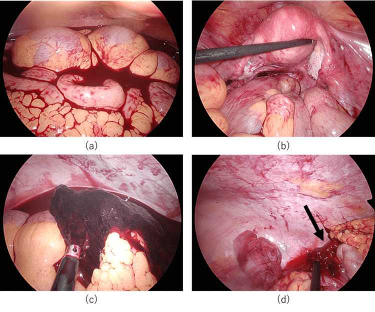
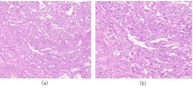
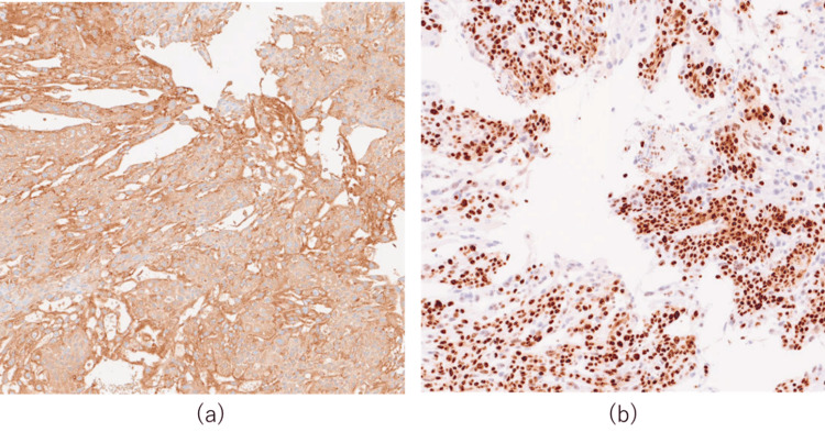
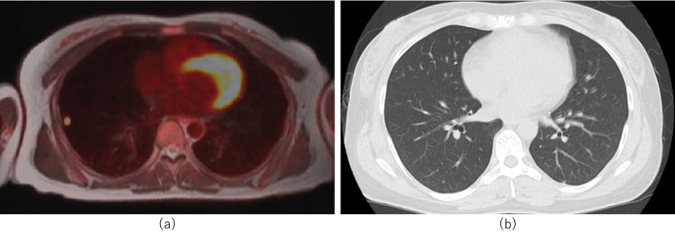
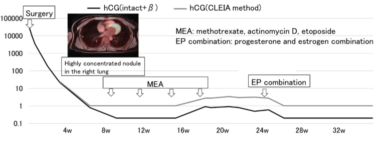
Extrauterine choriocarcinoma is uncommon and may be mistaken for a ruptured ectopic pregnancy. Rapid diagnosis is essential, as massive intraperitoneal bleeding can be fatal. Once histologically diagnosed, choriocarcinoma is highly chemosensitive. This case describes a 38-year-old woman who presented with sudden lower abdominal pain. Six weeks after her last menstrual period, serum human chorionic gonadotropin (hCG) was 28,834 mIU/mL. No intra-uterine gestational sac was found, and intraperitoneal bleeding was observed, suggesting an ectopic pregnancy. An emergency laparoscopic surgery revealed a blood clot and active bleeding on the peritoneal surface near the ileocecal region, which was resected. Histology revealed sheets of syncytiotrophoblasts and intermediate trophoblast cells without villi, and immunohistochemistry was diffusely positive for Ki-67 and hCG, confirming primary peritoneal choriocarcinoma. Staging imaging revealed no other lesions. The patient received four cycles of MEA chemotherapy (methotrexate, etoposide, actinomycin D) at three-week intervals, resulting in sustained hCG normalization and no evidence of recurrence at follow-up. Primary peritoneal choriocarcinoma should be considered in the differential diagnosis of intraperitoneal bleeding during early pregnancy. Even when ectopic pregnancy is suspected, the excised tissue must be submitted for histopathological examination so that chemotherapy can be initiated promptly in case of choriocarcinoma.
Keywords: extrauterine choriocarcinoma; gestational trophoblastic neoplasia; laparoscopy; peritoneal pregnancy; pituitary hcg.
Copyright © 2025, Maekawa et al.
Conflict of interest statement
Human subjects: Informed consent for treatment and open access publication was obtained or waived by all participants in this study. Conflicts of interest: In compliance with the ICMJE uniform disclosure form, all authors declare the following: Payment/services info: All authors have declared that no financial support was received from any organization for the submitted work. Financial relationships: All authors have declared that they have no financial relationships at present or within the previous three years with any organizations that might have an interest in the submitted work. Other relationships: All authors have declared that there are no other relationships or activities that could appear to have influenced the submitted work.
Figures
References
-
- Epidemiology, diagnosis, and treatment of gestational trophoblastic disease: A Society of Gynecologic Oncology evidence-based review and recommendation. Horowitz NS, Eskander RN, Adelman MR, Burke W. Gynecol Oncol. 2021;163:605–613. - PubMed
-
- Japan Society of Gynecologic Oncology. Endometrial Cancer Treatment Guidelines 2023 Edition (In Japanese) Tokyo: Japan Society of Gynecologic Oncology (JSGO)Tokyo; 2023. Guidelines for treatment of uterine body neoplasm.
-
- Ki-67 labeling index in the differential diagnosis of exaggerated placental site, placental site trophoblastic tumor, and choriocarcinoma: a double immunohistochemical staining technique using Ki-67 and Mel-CAM antibodies. Ming Shih IE, Kurman RJ. Hum Pathol. 1998;29:27–33. - PubMed
Publication types
LinkOut - more resources
Full Text Sources