

Dynamic Multi-Image Weighting for Automated Detection and Diagnosis of Abnormal Urinary Tract on Voiding Cystourethrography with a Deep Learning System: A Retrospective, Large-Scale, Multicenter Study
- PMID: 40698328
- PMCID: PMC12280329
- DOI: 10.34133/research.0771
Dynamic Multi-Image Weighting for Automated Detection and Diagnosis of Abnormal Urinary Tract on Voiding Cystourethrography with a Deep Learning System: A Retrospective, Large-Scale, Multicenter Study
Abstract
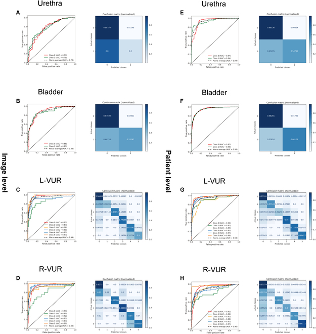
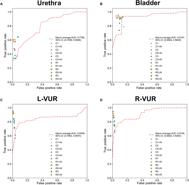
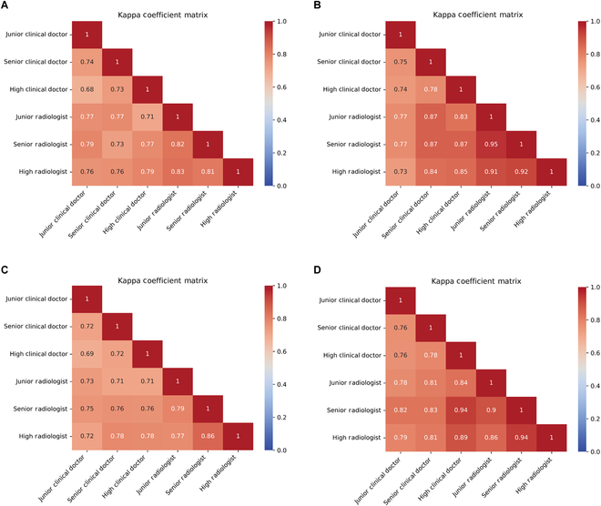
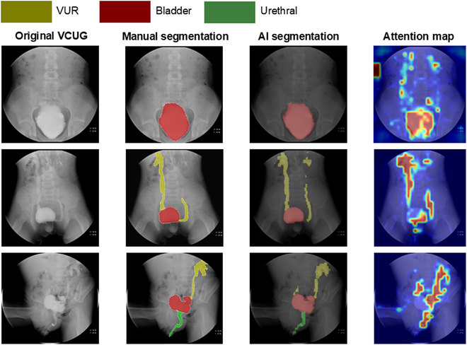
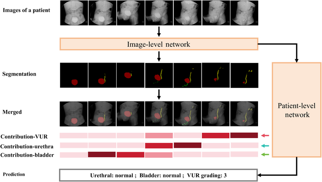
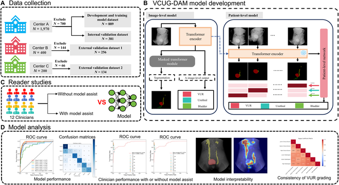
We aimed to develop a voiding cystourethrography (VCUG) diagnostic artificial intelligence model (VCUG-DAM), which relies on a novel architecture to automatically segment and diagnose the bladder, urethra, and ureters using a single VCUG image, while dynamically assessing the relative importance of each image. A total of 7,899 VCUG images from 1,660 patients across 15 Chinese hospitals were collected between 2021 and 2023. In stage 1, we assessed the performances of the VCUG-DAM model. The patient-level area under the curve (AUC) of VCUG-DAM was 0.8772, 0.7752, 0.9443, and 0.9342 for bladder, urethral, left, and right vesicoureteral reflux (VUR), respectively. In stage 2, we explored whether the VCUG-DAM model could improve the diagnostic ability of clinicians. VCUG-DAM improved the clinician's diagnostic performance, with mean AUCs increasing from 0.8185 to 0.9456 for the bladder, 0.6507 to 0.7943 for the urethra, 0.6288 to 0.9641 for the left VUR, and 0.7305 to 0.9506 for the right VUR (all P < 0.0001). In stage 3, the consistency of the VCUG-DAM for VUR grading was validated. VCUG-DAM improved inter-clinician agreement for VUR grading. The fully automated VCUG-DAM demonstrated high accuracy, reliability, and robustness in multitask diagnoses of urinary tract abnormalities across multiple VCUG images, while improving the diagnostic ability of clinicians as an auxiliary tool.
Copyright © 2025 Min Wu et al.
Conflict of interest statement
Competing interests: The authors declare that they have no competing interests.
Figures
Similar articles
-
Ultrasound and 99mTc-DMSA Scan Versus Voiding Cystourethrography in Diagnosis of Vesicoureteral Reflux in Children.Med J Islam Repub Iran. 2025 Mar 26;39:47. doi: 10.47176/mjiri.39.47. eCollection 2025. Med J Islam Repub Iran. 2025. PMID: 40740575 Free PMC article.
-
Dimercaptosuccinic acid scan or ultrasound in screening for vesicoureteral reflux among children with urinary tract infections.Cochrane Database Syst Rev. 2016 Jul 5;7(7):CD010657. doi: 10.1002/14651858.CD010657.pub2. Cochrane Database Syst Rev. 2016. PMID: 27378557 Free PMC article.
-
Radiation exposure in pediatric videourodynamics: An evaluation of safety in comparison to voiding cystourethrogram.J Pediatr Urol. 2024 Aug;20(4):745.e1-745.e6. doi: 10.1016/j.jpurol.2024.06.003. Epub 2024 Jun 7. J Pediatr Urol. 2024. PMID: 38908983
-
Voiding cystourethrography practices: experiences in a tertiary pediatric referral hospital.Acta Radiol. 2025 Jun 26:2841851251344466. doi: 10.1177/02841851251344466. Online ahead of print. Acta Radiol. 2025. PMID: 40569431
-
To screen or not to screen for vesicoureteral reflux in children with ureteropelvic junction obstruction: a systematic review.Eur J Pediatr. 2017 Jan;176(1):1-9. doi: 10.1007/s00431-016-2818-3. Epub 2016 Nov 25. Eur J Pediatr. 2017. PMID: 27888411
References
-
- Arlen AM, Amin J, Leong T. Voiding cystourethrogram: Who gets a cyclic study and does it matter? J Pediatr Urol. 2022;18(3):378–382. - PubMed
-
- Damasio MB, Donati F, Bruno C, Darge K, Mentzel HJ, Ključevšek D, Napolitano M, Ozcan HN, Riccabona M, Smets AM, et al. Update on imaging recommendations in paediatric uroradiology: The European Society of Paediatric Radiology workgroup session on voiding cystourethrography. Pediatr Radiol. 2024;54(4):606–619. - PubMed
-
- Lo WC, Wang CR, Lim KE. Diagnosis of the congenital urethral anomalies of male child by voiding cystourethrography. Acta Paediatr Taiwan. 1999;40(3):152–156. - PubMed
-
- Wang X, Chen HS, Wang C, Luo XG, Wang YX, Ye ZH, Liu X, Wei GH. A grading system for evaluation of bladder trabeculation. World J Urol. 2023;41(9):2443–2449. - PubMed
LinkOut - more resources
Full Text Sources
