. 2025 Jul 8:13:1587080.
doi: 10.3389/fbioe.2025.1587080. eCollection 2025.

Lung-resident SARS-CoV-2 peptide-specific immune responses in perfused 3D human lung explant models

Affiliations

Lung-resident SARS-CoV-2 peptide-specific immune responses in perfused 3D human lung explant models

Kayla F Goliwas et al. Front Bioeng Biotechnol. .

Abstract

Introduction: Multi-specific and long-lasting T-cell immunity has been recognized to indicate long-term protection against pathogens, including the novel coronavirus, SARS-CoV-2, which is the causative agent of the COVID-19 pandemic. Functional significance of peripheral memory T cells in individuals recovered from COVID-19 (COVID-19+) is beginning to be appreciated; however, the role of lung tissue-resident memory (lung TRM) T cells in SARS-CoV-2 infection is still being investigated. This is, in part, due to the lack of preclinical tissue models available to follow the convalescence period.

Methods: Here, we utilize a perfused three-dimensional (3D) human lung-tissue model and show pre-existing local T-cell immunity against SARS-CoV-2 proteins in lung tissues.

Results: We report ex vivo maintenance of functional multi-specific IFN-γ-secreting lung TRM T cells in COVID-19+ and their induction in lung tissues of vaccinated COVID-19+ subjects. Importantly, we identify SARS-CoV-2 peptide-responding memory B cells and IgA+ plasma cells in ex vivo cultured lung tissues of COVID-19+. Furthermore, lung tissue IgA levels were increased in COVID-19+ and responded to peptide stimulation.

Discussion: In our study, we highlight the importance of utilization of human lung-tissue models to understand the local antiviral immune response in the lung to protect against SARS-CoV-2 infection.

Keywords: COVID-19; SARS-CoV-2 infection; human lung-tissue model; local antiviral immune response; perfused lung explant.


Conflict of interest statement

The authors declare that the research was conducted in the absence of any commercial or financial relationships that could be construed as a potential conflict of interest.

Figures

FIGURE 1
FIGURE 1
Ex vivo perfusion culture of the human lung tissue and exposure to SARS-CoV-2 peptides. (A) Ex vivo model setup process. (B) Bioreactor chamber showing ECM volume-containing tissue. (C) Peptide exposure protocol.
FIGURE 2
FIGURE 2
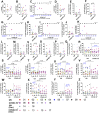
Local T-cell response to SARS-CoV-2 peptides within lung tissue cores. (A, B, D–F, H, J, and L–Q) Baseline T-cell differences within lung tissue from uninfected (UN) individuals and individuals recovered from COVID-19 (COVID-19+). (C, G, I, and K) Correlation between the convalescence period for COVID-19+ samples and the percentage of CD4+ T cells (C), PD-1+ CD8+ T cells (G), CD154+ CD69+ CD8+ T cells (J), and HLA-DR+ CD38+ CD8+ T cells (L). (R–Z) Impact of SARS-CoV-2 peptide exposure on T-cell populations in UN and COVID-19+ lung tissues (n = 10 UN and n = 8 COVID-19+ [mean (center line) ± SEM]). Statistics shown in blue are comparisons between control and peptide-exposed samples within each group (UN and COVID-19+). Statistics shown in black are the change in response between UN and COVID-19+ for each peptide when compared to the corresponding control.
FIGURE 3
FIGURE 3
Memory T-cell response to SARS-CoV-2 peptides within the lung. (A, C, E–G, I, K, and L) Baseline differences in memory T cells within lung tissue from uninfected (UN) individuals and individuals recovering from COVID-19 (COVID-19+). (B, D, H, J, and M) Correlation between the convalescence period for COVID-19+ samples and the percentage of CD4+ tissue-resident memory (TRM) T cells (B), CD8+ TRM T cells (D), TRM IFN-γ+ CD4+ T cells (H), TRM IFN-γ+ CD8+ T cells (J), and effector-memory CD8+ T cells (M). (N–U) Impact of SARS-CoV-2 peptide exposure on memory T-cell populations in UN and COVID-19+ lung tissues (n = 10 UN and n = 8 COVID-19+ [mean (center line) ± SEM]). Statistics shown in blue are comparisons between control and peptide-exposed samples within each group (UN and COVID-19+). Statistics shown in black are the change in the response between UN and COVID-19+ for each peptide when compared to the corresponding control.
FIGURE 4
FIGURE 4
Local B-cell response to SARS-CoV-2 peptides within the lung tissues. (A) Baseline differences in B-cell populations within lung tissue from uninfected (UN) individuals and individuals recovering from COVID-19 (COVID-19+). (B) Correlation between the convalescence period for COVID-19+ samples and the percentage of CD19+ B cells. (C–E) Impact of SARS-CoV-2 peptide exposure on B-cell populations in UN and COVID-19+ lung tissues. (F) Correlation between the convalescence period for COVID-19+ samples and the percentage of plasma cells. (G) Quantification of circulating SARS-CoV-2-specific IgA; opaque symbols indicate positive samples (measurements over threshold). (H). Photomicrographs showing CD138+ plasma cells (red) secreting IgA (green) in UN (left) and COVID-19+ samples (right) at day 0 (starting tissue). White arrows pointing to CD138+ plasma cells secreting IgA, gray dashed arrows pointing to CD138+ plasma cells without IgA secretion. (I and J). Correlation table showing correlation among the convalescence period, peptide response, and class-switched memory B populations (I) and the convalescence period, peptide response, and IgA secretion (J) (n = 5–10 UN and n = 6–8 COVID-19+ [mean (center line) ± SEM]). Statistics shown in blue are comparisons between control and peptide-stimulated samples within each group (UN and COVID-19+). Statistics shown in black are the change in response between UN and COVID-19+ for each peptide when compared to the corresponding controls.

Similar articles

  • Local SARS-CoV-2 Peptide-Specific Immune Responses in Lungs of Convalescent and Uninfected Human Subjects.
    Goliwas KF, Wood AM, Simmons CS, Khan R, Khan SA, Wang Y, Berry JL, Athar M, Mobley JA, Kim YI, Thannickal VJ, Harrod KS, Donahue JM, Deshane JS. Goliwas KF, et al. medRxiv [Preprint]. 2022 Jan 21:2021.09.02.21263042. doi: 10.1101/2021.09.02.21263042. medRxiv. 2022. PMID: 34518842 Free PMC article. Preprint.
  • Type I and Type III Interferons Restrict SARS-CoV-2 Infection of Human Airway Epithelial Cultures.
    Vanderheiden A, Ralfs P, Chirkova T, Upadhyay AA, Zimmerman MG, Bedoya S, Aoued H, Tharp GM, Pellegrini KL, Manfredi C, Sorscher E, Mainou B, Lobby JL, Kohlmeier JE, Lowen AC, Shi PY, Menachery VD, Anderson LJ, Grakoui A, Bosinger SE, Suthar MS. Vanderheiden A, et al. J Virol. 2020 Sep 15;94(19):e00985-20. doi: 10.1128/JVI.00985-20. Print 2020 Sep 15. J Virol. 2020. PMID: 32699094 Free PMC article.
  • Antibody tests for identification of current and past infection with SARS-CoV-2.
    Fox T, Geppert J, Dinnes J, Scandrett K, Bigio J, Sulis G, Hettiarachchi D, Mathangasinghe Y, Weeratunga P, Wickramasinghe D, Bergman H, Buckley BS, Probyn K, Sguassero Y, Davenport C, Cunningham J, Dittrich S, Emperador D, Hooft L, Leeflang MM, McInnes MD, Spijker R, Struyf T, Van den Bruel A, Verbakel JY, Takwoingi Y, Taylor-Phillips S, Deeks JJ; Cochrane COVID-19 Diagnostic Test Accuracy Group. Fox T, et al. Cochrane Database Syst Rev. 2022 Nov 17;11(11):CD013652. doi: 10.1002/14651858.CD013652.pub2. Cochrane Database Syst Rev. 2022. PMID: 36394900 Free PMC article.
  • Detectable SARS-CoV-2 specific immune responses in recovered unvaccinated individuals 250 days post wild type infection.
    Weigl N, Pleimelding C, Gilberg L, Huynh D, Brand I, Bruger J, Frese J, Eser TM, Ahmed MIM, Guggenbuehl-Noller JM, Stirner R, Hoelscher M, Pritsch M, Geldmacher C, Kobold S, Roider J; KoCo19-/ORCHESTRA-study group. Weigl N, et al. PLoS One. 2025 Jun 11;20(6):e0325923. doi: 10.1371/journal.pone.0325923. eCollection 2025. PLoS One. 2025. PMID: 40498720 Free PMC article.
  • Rapid, point-of-care antigen tests for diagnosis of SARS-CoV-2 infection.
    Dinnes J, Sharma P, Berhane S, van Wyk SS, Nyaaba N, Domen J, Taylor M, Cunningham J, Davenport C, Dittrich S, Emperador D, Hooft L, Leeflang MM, McInnes MD, Spijker R, Verbakel JY, Takwoingi Y, Taylor-Phillips S, Van den Bruel A, Deeks JJ; Cochrane COVID-19 Diagnostic Test Accuracy Group. Dinnes J, et al. Cochrane Database Syst Rev. 2022 Jul 22;7(7):CD013705. doi: 10.1002/14651858.CD013705.pub3. Cochrane Database Syst Rev. 2022. PMID: 35866452 Free PMC article.

References

    1. Abbasi J. (2021). SARS-CoV-2 variant antibodies wane 6 Months after vaccination. JAMA 326, 901. 10.1001/jama.2021.15115 - DOI - PubMed
    1. Akkaya M., Kwak K., Pierce S. K. (2020). B cell memory: building two walls of protection against pathogens. Nat. Rev. Immunol. 20, 229–238. 10.1038/s41577-019-0244-2 - DOI - PMC - PubMed
    1. Biospace (2020). Enzo announces issuance of U.S. Patent for methods of using proprietary compound SK1-I in patients; exploring options for development as a potential treatment for COVID-19. Farmingdale, NY: Enzo Biochem, Inc. Available online at: https://www.biospace.com/article/releases/enzo-announces-issuance-of-u-s....
    1. Boppana S., Qin K., Files J. K., Russell R. M., Stoltz R., Bibollet-Ruche F., et al. (2021). SARS-CoV-2-specific circulating T follicular helper cells correlate with neutralizing antibodies and increase during early convalescence. PLoS Pathog. 17, e1009761. 10.1371/journal.ppat.1009761 - DOI - PMC - PubMed
    1. Cagigi A., Yu M., Österberg B., Svensson J., Falck-Jones S., Vangeti S., et al. (2021). Airway antibodies emerge according to COVID-19 severity and wane rapidly but reappear after SARS-CoV-2 vaccination. JCI Insight 6, e151463. 10.1172/jci.insight.151463 - DOI - PMC - PubMed

LinkOut - more resources