. 2025 Sep 2;13(9):e0080925.
doi: 10.1128/spectrum.00809-25. Epub 2025 Jul 23.

Hepatitis B surface antigen is upregulated by HIV Tat in an HIV-hepatitis B virus co-infection model system

Affiliations

Hepatitis B surface antigen is upregulated by HIV Tat in an HIV-hepatitis B virus co-infection model system

Wei Zhao et al. Microbiol Spectr. .

Abstract

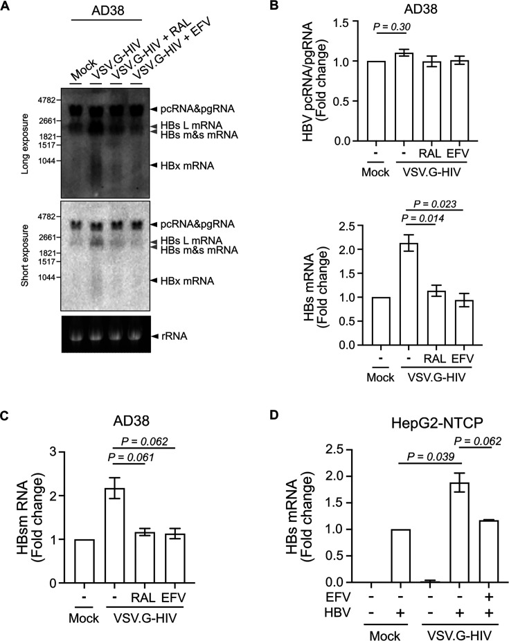
People with human immunodeficiency virus-hepatitis B virus (HIV-HBV) co-infection have faster rates of liver disease progression and an increase in hepatocellular carcinoma compared to people with HBV mono-infection. Given that HIV can infect multiple cells in the liver, including hepatocytes, we hypothesized that HIV will impact HBV replication through HIV viral proteins that can impact HBV replication either directly or indirectly, via effects on cellular pathways. Following infection of sodium taurocholate co-transporting polypeptide (NTCP)-expressing HepG2 cells with HBV and vesicular stomatitis virus G protein (VSV.G)-pseudotyped HIV, we found that productive HIV infection led to a twofold upregulation of HBV surface (HBs) mRNA and a marked increase in intracellular production and cellular retention of HBs antigen (HBsAg). Overexpression of HIV Tat protein, but not other HIV proteins, by DNA plasmid transfection in the HBV-producing cell line AD38 significantly stimulated HBs mRNA expression. This could be rescued by CDK9 inhibition with BAY-1251152. This study provides new insights into the mechanisms by which HIV directly impacts HBV replication and has implications for understanding adverse liver outcomes in people living with HIV and HBV.IMPORTANCEPeople with both human immunodeficiency virus (HIV) and hepatitis B virus (HBV) face faster liver disease progression and a higher risk of liver cancer than those with HBV alone. This study investigated how HIV affects HBV replication in liver cells and found that HIV infection increases the production of a key HBV surface protein (HBsAg) by enhancing the expression of its gene (HBs). This effect is driven by the HIV Tat protein. Notably, blocking the CDK9 pathway prevented this increase, suggesting a possible explanation for the adverse liver outcomes in co-infected individuals. Our findings have implications for interventions aiming to cure HIV through latency reversal, as these interventions can specifically increase the Tat protein. Future exploratory treatment strategies, such as Tat inhibitors, could play a role in the management of people with HIV and HBV at high risk of liver disease.

Keywords: CDK9; HBsAg; Tat; co-infection; hepatitis B virus; hepatocyte; human immunodeficiency virus; liver.


Conflict of interest statement

S.R.L. has received funding from the National Health and Medical Research Council of Australia (NHMRC), the National Institutes of Health (NIH), amfAR, The Australian Centre for HIV and Hepatitis Research, Melbourne HIV Cure Consortium, Gilead Sciences, Merck, ViiV, Abbvie, and Gilead outside the submitted work. F.J.R. receives institutional support as a coinvestigator and is subcontracted by the Peter MacCallum Cancer Centre for an investigator-initiated trial, which receives funding support from Sanofi/Regeneron Pharmaceuticals.

Figures

Fig 1
Fig 1
High levels of HIV-HBV co-infection in AD38 and HepG2-NTCP cells using VSV.G pseudotyped HIV virus. (A) Percentage of AD38 cells expressing green fluorescent protein (GFP) 5 days post VSV.G-pseudotyped HIV infection. Cells were treated with or without 10 µM raltegravir (RAL) or 300 nM efavirenz (EFV) 24 h before and immediately after HIV infection. GFP expression was determined by flow cytometry (N = 3). (B) Frequency of HIV integration in AD38 cells 5 days post VSV.G-pseudotyped HIV infection with or without RAL/EFV treatment. HIV integration was measured by real-time PCR for Alu-LTR and normalized to CCR5 copy number. The detection limit for the Alu-LTR was 200 copies/106 cells and is shown as a dashed line (N = 3). (C) GFP and HBsAg expression in mock, HBV-mono, HIV-mono, and HIV-HBV co-infected HepG2-NTCP cells. HepG2-NTCP cells were infected with HBV inoculum derived from AD38 cells for 10 days and VSV.G-pseudotyped HIV virus for another 5 days. Cells were immunostained for GFP (green) and HBsAg (red). DNA was counterstained with DAPI (blue). Bars, 10 µm. Results are representative of at least three experiments. (D) Percentage of HepG2-NTCP cells expressing GFP following HBV and VSV.G-pseudotyped HIV infection. Cells were treated with or without 300 nM EFV 24 h before and after HIV infection (N = 3). (E) HIV integration in HepG2-NTCP cells following HBV and VSV.G-pseudotyped HIV infection with or without EFV treatment (N = 4). In all graphs, the horizontal bar represents the mean.
Fig 2
Fig 2
Productive HIV infection leads to an increase in intracellular HBsAg in AD38 and HepG2-NTCP cells. (A) Western blot with anti-HBV PreS2 and anti-GAPDH antibodies using lysates from AD38 cells 5 days post-infection with VSV.G-pseudotyped HIV treated with or without 10 µM RAL or 300 nM EFV 24 h before and after HIV infection. Results are representative of at least three experiments. (B) Western blot with anti-HBV PreS2 and anti-GAPDH antibodies using lysates from HepG2-NTCP cells infected with HBV, VSV.G-pseudotyped HIV infection or both and treated with or without 300 nM EFV 24 h before and immediately after HIV infection. Results are representative of at least three experiments. Results are representative of at least three experiments. (C) HIV integration in AD38 cells with or without productive HIV infection. GFP+ and GFP− cells were collected by flow sorting 5 days post VSV.G-pseudotyped HIV infection. HIV integration was quantified using real-time PCR for Alu-LTR and normalized to CCR5 copy numbers as a housekeeping gene. The detection limit for the Alu-LTR was 200 copies/106 cells and is shown as a dashed line (N = 2). (D) Representative example (from two separate experiments) of intracellular HBsAg levels in AD38 cells with or without productive HIV infection. GFP+ and GFP− cells were collected by flow sorting 5 days post VSV.G-pseudotyped HIV infection. Cell lysates of unsorted and sorted samples were examined by Western blot with anti-HBV PreS2 and anti-GAPDH antibodies. (E) HIV integration in AD38 cells infected with an HIV virus expressing GFP or a luciferase (luc) reporter. Cells were treated with or without 300 nM EFV 24 h before and immediately after HIV infection (N = 2). (F) Representative example (from two independent experiments) of intracellular HBsAg levels in AD38 cells 5 days post VSV.G-pseudotyped HIV infection expressing GFP or a luciferase (luc) reporter. Cells were treated with or without 300 nM EFV 24 h before and immediately after HIV infection. AD38 cell lysates were examined by Western blot with anti-HBV PreS2 and anti-GAPDH antibodies. In all graphs, the horizontal bar represents the mean.
Fig 3
Fig 3
Productive HIV infection leads to a twofold increase in HBs mRNA in AD38 and HepG2-NTCP cells. (A) Northern blot of RNA extracted from AD38 cells 5 days post VSV.G-pseudotyped HIV infection. Cells were treated with or without 10 µM RAL or 300 nM EFV 24 h before and immediately after HIV infection. A genomic length HBV-DNA probe or a ribosomal probe was used. A long (upper panel) and short (lower panel) exposure time is shown. Arrows in black indicate pcRNA/pgRNA, HBx mRNA or ribosomal RNA. Arrows in gray indicate large (L), medium (M), and small (S) HBs mRNA; Representative example from three separate experiments. (B) Fold change of HBV pcRNA/pgRNA and HBs mRNA on Northern blot (A) using image density (N = 3). (C) Fold change of HBs mRNA expression in AD38 cells 5 days post VSV.G-pseudotyped HIV infection. Cells were treated with or without 10 µM RAL or 300 nM EFV 24 h before and after HIV infection. HBs mRNA was quantified by real-time PCR using two sets of specific primers, which quantified either pcRNA/pgRNA (3.5 kb) only or together with HBs mRNAs (3.5 kb + 2.4 kb + 2.1 kb). HBV PreC (pcRNA/pgRNA) was subtracted from PreC and S (pcRNA/pgRNA with HBs mRNAs) and normalized to the expression of the housekeeping gene RPLP0 (N = 3). (D) Fold change of HBs mRNA level in HepG2-NTCP cells following HBV and VSV.G-pseudotyped HIV infection. Cells were treated with or without 300 nM EFV 24 h before and after HIV infection. HBs mRNA was quantified by real-time PCR and normalized to the expression of the housekeeping gene RPLP0 (N = 3). In all graphs, the columns and error bars represent mean and SEM.
Fig 4
Fig 4
HIV Tat upregulates the transcription level of HBs via CDK9/cyclin T2. (A) Fold change of HBs mRNA in AD38 cells following transfection of plasmids expressing either HIV NL4-3-GFP or Gag-GFP (left panel) or FLAG-tagged Nef, Rev, Tat, Vpr, or Vpu (right panel). The cells were transfected with plasmid DNA and enriched following flow sorting, based on expression of either GFP expression or FLAG intracellular staining. HBs mRNA was quantified by real-time PCR and normalized to the expression of the housekeeping gene RPLP0 (N = 5). (B) Western blot with anti-HBV PreS2 and anti-α-tubulin antibodies using lysates from AD38 cells 2 days post-transfection with lipid nanoparticles (LNPs), which were empty or co-formulated with Tat mRNA (Tat-LNP) ranging from 50 to 400 ng/mL. Results are representative of three experiments. (C) Fold change of HBs mRNA level in AD38 cells 2 days post Tat-LNP transfection. HBs mRNA was quantified by real-time PCR and normalized to the expression of the housekeeping gene RPLP0 (N = 3). (D) Western blot with anti-RNA-Pol2-S2p, anti-HBV PreS2, and anti-α-tubulin antibodies. AD38 cells were transfected with or without 200 ng/mL Tat-LNP for 48 h and treated with or without 1 µM BAY1251152 for another 16 h. Results are representative of two experiments. (E) Fold change of HBs mRNA level in AD38 cells transfected with or without 200 ng/mL Tat-LNP for 48 h followed by 1 µM BAY1251152 treatment for another 16 h. HBs mRNA was quantified by real-time PCR and normalized to the expression of the housekeeping gene RPLP0 (N = 2). In all graphs, the columns and error bars represent mean and SEM.

Similar articles

References

    1. Platt L, French CE, McGowan CR, Sabin K, Gower E, Trickey A, McDonald B, Ong J, Stone J, Easterbrook P, Vickerman P. 2020. Prevalence and burden of HBV co-infection among people living with HIV: a global systematic review and meta-analysis. J Viral Hepat 27:294–315. doi: 10.1111/jvh.13217 - DOI - PMC - PubMed
    1. Smith CJ, Ryom L, Weber R, Morlat P, Pradier C, Reiss P, de Wit S, Law M. 2014. Trends in underlying causes of death in people with HIV from 1999 to 2011 (D:A:D): a multicohort collaboration. Lancet 384:241–248. doi: 10.1016/S0140-6736(14)60604-8 - DOI - PubMed
    1. Boyd A, Gozlan J, Maylin S, Delaugerre C, Peytavin G, Girard PM, Zoulim F, Lacombe K. 2014. Persistent viremia in human immunodeficiency virus/hepatitis B coinfected patients undergoing long-term tenofovir: virological and clinical implications. Hepatology 60:497–507. doi: 10.1002/hep.27182 - DOI - PubMed
    1. Klein MB, Althoff KN, Jing Y, Lau B, Kitahata M, Lo Re V 3rd, Kirk GD, Hull M, Kim HN, Sebastiani G, et al. 2016. Risk of end-stage liver disease in HIV-viral hepatitis coinfected persons in North America from the early to modern antiretroviral therapy eras. Clin Infect Dis 63:1160–1167. doi: 10.1093/cid/ciw531 - DOI - PMC - PubMed
    1. Dezanet LNC, Kassime R, Miailhes P, Lascoux-Combe C, Chas J, Maylin S, Gabassi A, Rougier H, Delaugerre C, Lacombe K, Boyd A. 2021. Effect of viral replication and liver fibrosis on all-cause mortality in HIV/HBV coinfected individuals: a retrospective analysis of a 15-year longitudinal cohort. MedRxiv. doi: 10.1101/2021.04.13.21255432 - DOI - PubMed

MeSH terms

Substances

LinkOut - more resources