

Balancing Microglial Density and Activation in Central Nervous System Development and Disease
- PMID: 40699743
- PMCID: PMC12109793
- DOI: 10.3390/cimb47050344
Balancing Microglial Density and Activation in Central Nervous System Development and Disease
Abstract
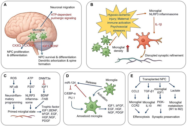
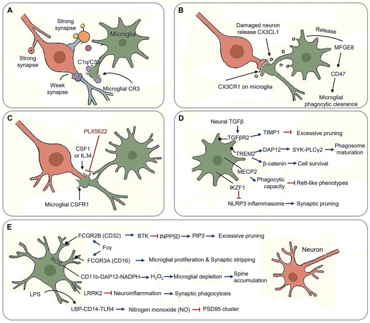
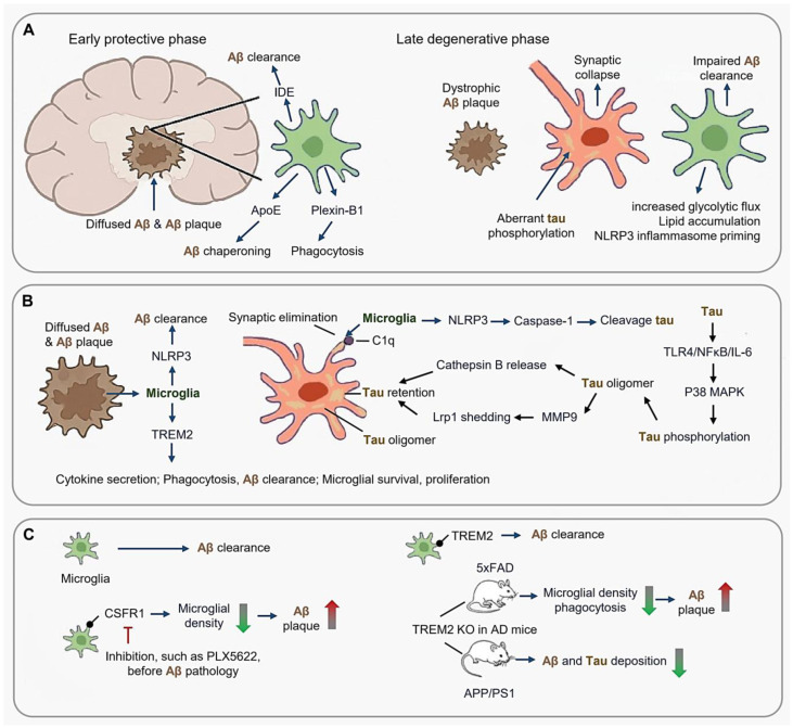
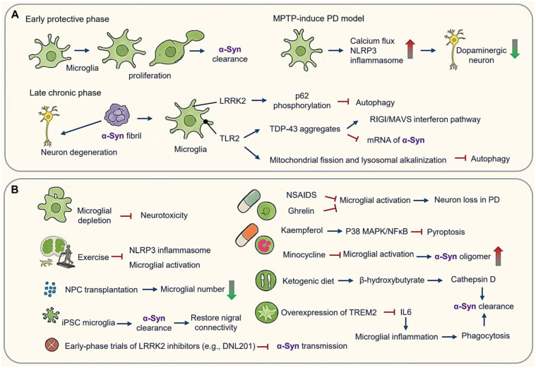
Microglia, the resident immune cells of the central nervous system, play multifaceted roles in both health and disease. During development, they regulate neurogenesis and refine neural circuits through synaptic pruning. In adulthood, microglia maintain homeostasis and dynamically respond to pathological insults, where they contribute to responding to neuroinflammatory challenges. This review summarizes microglial contributions to neurodevelopment and also outlines their function across various neurodegenerative diseases, such as Alzheimer's disease, Parkinson's disease, Huntington's disease, and amyotrophic lateral sclerosis, highlighting both protective and detrimental effects. Finally, recent advances in microglial-targeted therapies and lifestyle-based interventions are highlighted, underscoring the translational potential of modulating microglial states. Elucidating the dual roles of microglia in development and disease could guide the design of therapeutic strategies aimed at enhancing neuroprotection while minimizing neurotoxicity.
Keywords: lifestyle modifications; microglia; neurogenesis; neurological diseases; synaptic pruning; therapeutic interventions.
Conflict of interest statement
The authors have no conflicts of interest to disclose.
Figures
Similar articles
-
Decoding the Dialogue: Immunity and central nervous system interactions in neurodegenerative diseases.Egypt J Immunol. 2025 Jul;32(3):20-31. doi: 10.55133/eji.320303. Egypt J Immunol. 2025. PMID: 40682794 Review.
-
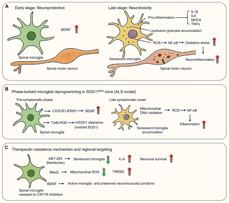
Emerging roles of microglia and neuroinflammation in Huntington's disease: From pathophysiology to clinical trials.J Huntingtons Dis. 2025 Aug;14(3):241-257. doi: 10.1177/18796397251330144. Epub 2025 Mar 28. J Huntingtons Dis. 2025. PMID: 40152279 Review.
-
Microglial activation as a hallmark of neuroinflammation in Alzheimer's disease.Metab Brain Dis. 2025 May 17;40(5):207. doi: 10.1007/s11011-025-01631-9. Metab Brain Dis. 2025. PMID: 40381069 Review.
-
Glucose Metabolic Reprogramming in Microglia: Implications for Neurodegenerative Diseases and Targeted Therapy.Mol Neurobiol. 2025 Jul;62(7):8204-8221. doi: 10.1007/s12035-025-04775-y. Epub 2025 Feb 22. Mol Neurobiol. 2025. PMID: 39987285 Review.
-
Mesenchymal Stem Cell-Derived Extracellular Vesicles: Emerging Therapies for Neurodegenerative Diseases.Int J Nanomedicine. 2025 Jul 2;20:8547-8565. doi: 10.2147/IJN.S526945. eCollection 2025. Int J Nanomedicine. 2025. PMID: 40620684 Free PMC article. Review.
References
-
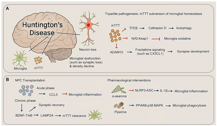
- Gunner G., Cheadle L., Johnson K.M., Ayata P., Badimon A., Mondo E., Nagy M.A., Liu L., Bemiller S.M., Kim K.W., et al. Sensory lesioning induces microglial synapse elimination via ADAM10 and fractalkine signaling. Nat. Neurosci. 2019;22:1075–1088. doi: 10.1038/s41593-019-0419-y. - DOI - PMC - PubMed
-
- Bennett M.L., Bennett F.C., Liddelow S.A., Ajami B., Zamanian J.L., Fernhoff N.B., Mulinyawe S.B., Bohlen C.J., Adil A., Tucker A., et al. New tools for studying microglia in the mouse and human CNS. Proc. Natl. Acad. Sci. USA. 2016;113:E1738–E1746. doi: 10.1073/pnas.1525528113. - DOI - PMC - PubMed
Publication types
Grants and funding
LinkOut - more resources
Full Text Sources
