. 2025 May 9;47(5):345.
doi: 10.3390/cimb47050345.

The N-Linked Glycosylation Site N201 in eel Lutropin/Choriogonadotropin Receptor Is Uniquely Indispensable for cAMP Responsiveness and Receptor Surface Loss, but Not pERK1/2 Activity

Affiliations

The N-Linked Glycosylation Site N201 in eel Lutropin/Choriogonadotropin Receptor Is Uniquely Indispensable for cAMP Responsiveness and Receptor Surface Loss, but Not pERK1/2 Activity

Munkhzaya Byambaragchaa et al. Curr Issues Mol Biol. .

Abstract

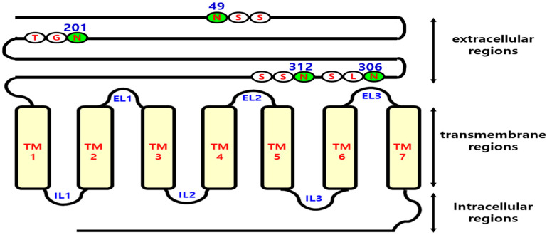
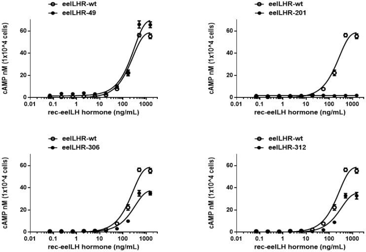
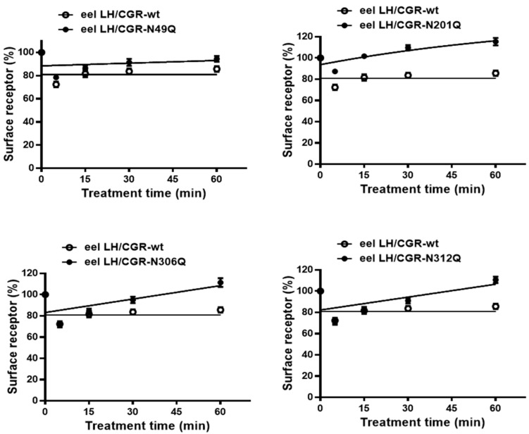
The seven transmembrane-spanning lutropin/chorionic gonadotropin receptors (LH/CGRs) trigger extracellular signal-related kinases (ERK1/2) via a noticeable network dependent on either G protein (Gαs) or β-arrestins. LH/CGRs are highly conserved, with the largest region within the transmembrane helices and common N-glycosylation sites in the extracellular domain. We aimed to determine the glycosylation sites that play crucial roles in cAMP and pERK1/2 regulation by constructing four mutants (N49Q, N201Q, N306Q, and N312Q). The cAMP response in cells expressing the N201Q mutant was completely impaired, despite high-dose agonist treatment. The cell-surface expression level was lowest in transiently transfected cells, but normal surface loss of the receptor occurred in cells expressing the wild-type and other mutant proteins. However, the N201Q mutant was only slightly reduced after 5 min of agonist stimulation. All mutants showed a peak in cAMP signaling 5 min after stimulation with a pERK1/2 agonist. Of note, cAMP activity was completely impaired in the N201Q mutant; however, this mutant still displayed a pERK1/2 response. These data show that the specific N-linked glycosylation site in eel LH/CGR is clearly distinguished by its differential responsiveness to cAMP signaling and pERK1/2 activity. Thus, we suggest that the cAMP and pERK1/2 signaling pathways involving eel LH/CGRs represent pleiotropic signal transduction induced by agonist treatment.

Keywords: N-linked glycosylation; cAMP responsive; eel LH/CGR; pERK1/2 activity.


Conflict of interest statement

The authors declare no conflicts of interest.

Figures

Figure 1
Figure 1
Schematic representation of the structure of wild-type eel LH/CGR and its N-linked glycosylation sites. Similar to the situation in mammals, N-linked glycosylation sites are present in the extracellular region of eel lutropin/chorionic gonadotropin receptor (LH/CGR). The extracellular domain of eel LH/CGR contains four putative N-linked glycosylation sites. Residue 49 is located in the anterior region of the N-terminus, whereas residue 201 is genetically conserved in both mammals and eels. Residues 306 and 312 are encoded in exon 10. When ligand stimulation induces phosphorylation of the C-terminal region of these receptors by GRKs, intracellular signal transduction is initiated. Therefore, signal transduction was analyzed in relation to variants at the glycosylation sites.
Figure 2
Figure 2
Cell-surface expression of eel LH/CGRs after transient transfection in HEK 293 cells. Wild-type eel LH/CGRs (LH/CGR-wt) and mutants were transiently expressed in HEK 293 cells, and their expression levels were determined using enzyme-linked immunosorbent assay (ELISA). Values are presented as the mean ± standard error of the mean from three independent experiments and normalized to the eel LH/CGR-wt level, which was set at 100%. Statistically significant differences were determined using one-way analysis of variance (ANOVA) followed by Tukey’s multiple comparisons test (* p < 0.05, ** p < 0.01).
Figure 3
Figure 3
Total cAMP levels in CHO-K1 cells transfected with eel LH/CGR-wt and N-linked glycosylation site mutants. After transfection in a 6-well plate, 10,000 cells were seeded in a 384-well plate for 48 h. The cells were then incubated with rec-eel LH for 30 min, and cAMP levels were analyzed using a homogeneous time-resolved fluorescence (HTRF) assay. The ratios of the values measured at 665 and 620 nm were calculated to determine the delta F% value. Using GraphPad Prism software 6.0, these data were then compared to the cAMP standard to determine the amount of cAMP produced. Data represent the mean ± standard error of the mean of three independent experiments. Mean values were fitted to a single-phase exponential decay curve.
Figure 4
Figure 4
Rmax value of cAMP for eel LH/CGR-wt and N-linked glycosylation mutants. The maximum cAMP values presented in Figure 3 and Table 1 are presented as bar graphs. Statistically significant differences were determined using one-way ANOVA, followed by Tukey’s multiple comparisons test (* p < 0.05, ** p < 0.01).
Figure 5
Figure 5
Time-dependent cell-surface loss in eel LH/CGR-wt and N-linked glycosylation mutants. The cell-surface expression level in the absence of agonist treatment was used as the baseline and set to 100% for comparison. The mean data were fitted to a single-phase exponential decay equation. The blank circles indicate the same curves for eel LH/CGR-wt.
Figure 6
Figure 6
Cell-surface loss of eel LH/CGR-wt and N-linked glycosylation mutants. Receptor surface loss is expressed as a percentage, with the absence of agonist treatment considered as 0% cell-surface loss. The decrease in cell-surface expression after 15 min of recombinant LH agonist treatment is presented as a percentage. Statistically significant differences were determined using one-way ANOVA, followed by Tukey’s multiple comparisons test (* p < 0.01).
Figure 7
Figure 7
Homogeneous time-resolved fluorescence analysis of pERK1/2 activity, presenting the basal signal and the increased activity 5 min after agonist treatment. The ratio of the values measured at 665 and 620 nm was calculated, with each time-0 result presented as the basal signal. (A) Presentation of the basal signal of pERK1/2. (B) Increase at 5 min compared with the basal level for each. * p < 0.05, indicating a significant difference between groups.
Figure 8
Figure 8
Homogeneous time-resolved fluorescence analysis results of pERK1/2 activity following treatment with recombinant eel LH for different times. All mutants were analyzed by comparing their measurements to the time-0 value before agonist treatment of eel LH/CGR-wt. The fold change values were calculated by setting the eel LH/CGR-wt time-0 value to 1, and they are presented as a graph. All mutants and LH/CGR-wt showed a sharp increase, reaching a peak value at 5 min, followed by a gradual decrease.
Figure 9
Figure 9
HpERK1/2 activation in HEK 293 cells transfected with eel LH/CGR-wt and mutants after stimulation with recombinant eel LH agonist. After transfection of HEK 293 cells with each plasmid, the cells were serum-starved for approximately 6 h before stimulation with 250 ng/mL agonist for the indicated time periods. Whole-cell lysates were isolated and analyzed for pERK1/2 and total ERK levels. (A) Western blotting results for pERK1/2 and total ERK levels. (B) Quantified pERK1/2 levels normalized to total ERK levels are expressed as a percentage of the maximal response, which was set to 100% for eel LH/CGR-wt at 5 min. Densitometry was performed to quantify the pERK1/2 bands.

Similar articles

References

    1. Shiraishi K., Ascoli M. Lutropin/choriogonadotropin stimulate the proliferation of primary cultures of rat Leydig cells through a pathway that involves activation of the extracellularly regulated kinase 1/2 cascade. Endocrinology. 2007;148:3214–3225. doi: 10.1210/en.2007-0160. - DOI - PMC - PubMed
    1. Ji I., Slaughter R.G., Ji T.H. N-linked oligosaccharides are not required for hormone binding of the lutropin receptor in a Leydig tumor cell line and rat granulosa cells. Endocrinology. 1990;127:494–496. doi: 10.1210/endo-127-1-494. - DOI - PubMed
    1. Ascoli M. Potential Leydig cell mitogenic signals generated by the wild-type and constitutively active mutants of the lutropin/choriogonadotropin receptor (LHR) Mol. Cell. Endocrinol. 2007;260–262:244–248. doi: 10.1016/j.mce.2005.09.016. - DOI - PMC - PubMed
    1. Kara E., Crepieux P., Gauthier C., Martinat N., Piketty V., Guillou F., Reiter E. A phosphorylation cluster of five serine and threonine residues in the C-terminus of the follicle-stimulating hormone receptor is important for desensitization but not for β-arrestin-mediated ERK activation. Mol. Endocrinol. 2006;20:3014–3026. doi: 10.1210/me.2006-0098. - DOI - PubMed
    1. Luttrell L.M., Wang J., Plouffe B., Smith J.S., Yamani L., Kaur S., Jean-Charles P.Y., Gauthier C., Lee M.H., Pani B., et al. Manifold roles of β-arrestin in GPCR signaling elucidated with siRNA and CRISPR/Cas9. Sci. Signal. 2018;11:549. doi: 10.1126/scisignal.aat7650. - DOI - PMC - PubMed

LinkOut - more resources