Review
. 2025 Jun 19;47(6):475.
doi: 10.3390/cimb47060475.

Integrating Genomics and Molecular Biology in Understanding Peritoneal Adhesion

Affiliations
Review

Integrating Genomics and Molecular Biology in Understanding Peritoneal Adhesion

Mirela Lungu et al. Curr Issues Mol Biol. .

Abstract

Peritoneal adhesions following surgical injury remain a major clinical challenge, often resulting in severe complications, such as intestinal obstruction, chronic pain, and infertility. This review systematically integrates recent genomic and molecular biology insights into the pathogenesis of peritoneal adhesions, explicitly focusing on molecular pathways, including TGF-β signaling, COX-2-mediated inflammatory responses, fibrinolytic balance (tPA/PAI-1), angiogenesis pathways (VEGF, PDGF), and extracellular matrix remodeling (MMPs/TIMPs). Newly conducted transcriptomic and proteomic analyses highlight distinct changes in gene expression patterns in peritoneal fibroblasts during adhesion formation, pinpointing critical roles for integrins, cadherins, selectins, and immunoglobulin superfamily molecules. Recent studies indicate significant shifts in TGF-β isoforms expression, emphasizing isoform-specific impacts on fibrosis and scarring. These insights reveal substantial knowledge gaps, particularly the differential regulatory mechanisms involved in fibrosis versus normal reparative reperitonealization. Future therapeutic strategies could target these molecular pathways and inflammatory mediators to prevent or reduce adhesion formation. Further research into precise genetic markers and the exploration of targeted pharmacological interventions remain pivotal next steps in mitigating postoperative adhesion formation and improving clinical outcomes.

Keywords: adhesion; adhesion genes; peritoneum; peritonitis; reintervention.


Conflict of interest statement

The authors declare no conflicts of interest.

Figures

Figure 1
Figure 1
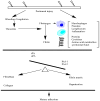
Peritoneal adhesion mechanism. Blue bands—mesothelium; circles—aggregated macrophages; brown lines—fibrin; star—myofibroblast to collagen fiber formation; and finally, mesothelium, red, blue, and yellow lines—artery, vein, and nerve, respectively. Adhesion development following surgery—preoperative overview of the peritoneal cavity. Major abdominal surgery is one example of a non-focal mesothelial injury that causes an uncontrollably large peritoneal macrophage aggregation (A) that acts as the nidus for the subsequent fibrin clot deposition (B). Fibrin deposition is promoted by coagulation and inflammation in concert. The intestine, for example, is currently affixed to the abdominal wall at anatomic (mesentery) and non-anatomic (adhesion) sites. Myofibroblasts, which migrate into the wound and fibrin clot to deposit extracellular matrix (ECM), such as collagen, are produced due to the mesenchymal to epithelial transition (C). The completion of adhesion development occurs when mesothelium covers the scar tissue. The lesion may become perfused and pain-sensitive due to blood vessels and nerves growing into it (D).
Figure 2
Figure 2
The peritoneal adhesion formation mechanism involves the fibrinogen pathway. tPA—tissue plasminogen activator, uPA—urokinase type plasminogen activator, PAI-1—plasminogen activator inhibitor 1, PAI-2—plasminogen activator inhibitor 2. Violet, blue, and black collared images: macrophage, lymphocyte, amino acid chains. When there is an imbalance between the plasminogen activator and plasminogen inhibitor factors, peritoneal adhesion is favored (as a result of collagen formation and organization).

References

    1. Capobianco A., Cottone L., Monno A., Manfredi A.A., Rovere-Querini P. The peritoneum: Healing, immunity, and diseases. J. Pathol. 2017;243:137–147. doi: 10.1002/path.4942. - DOI - PubMed
    1. Stratakis K., Kalogiannis E., Thanopoulos A., Grigoriadis G., Roditis S., Kaliatsi E.G., Nikiteas N., Kontzoglou K., Perrea D. Mechanisms and Therapeutic Approaches for Peritoneal Adhesions: A Comprehensive Review. Chirurgia. 2023;118:113–126. doi: 10.21614/chirurgia.2840. - DOI - PubMed
    1. Fometescu S.G., Costache M., Coveney A., Oprescu S.M., Serban D., Savlovschi C. Peritoneal fibrinolytic activity and adhesiogenesis. Chirurgia. 2013;108:331–340. - PubMed
    1. Van Baal J.O., Van de Vijver K.K., Nieuwland R., van Noorden C.J., van Driel W.J., Sturk A., Kenter G.G. The histophysiology and pathophysiology of the peritoneum. Tissue Cell. 2017;49:95–105. doi: 10.1016/j.tice.2016.11.004. - DOI - PubMed
    1. Trayhurn P. Hypoxia and adipose tissue function and dysfunction in obesity. Physiol. Rev. 2013;93:1–21. doi: 10.1152/physrev.00017.2012. - DOI - PubMed

LinkOut - more resources