. 2025 Jul 7;8(4):74.
doi: 10.3390/mps8040074.

A Framework for Corticomuscle Control Studies Using a Serious Gaming Approach

Affiliations

A Framework for Corticomuscle Control Studies Using a Serious Gaming Approach

Pedro Correia et al. Methods Protoc. .

Abstract

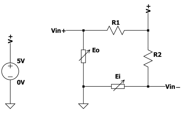
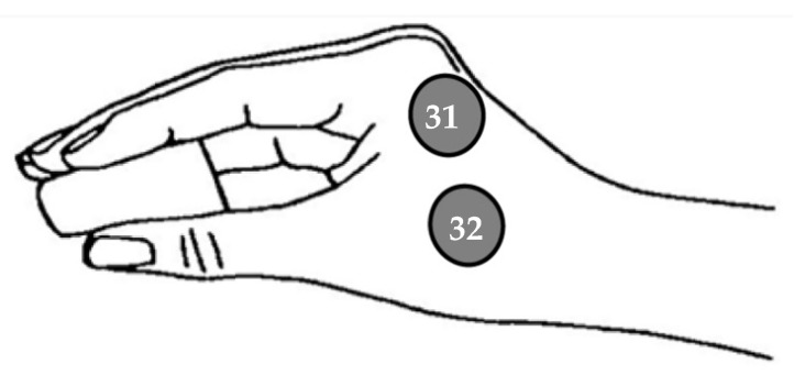
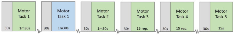
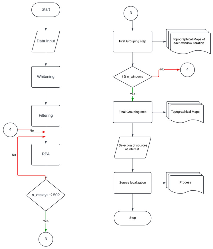
Sophisticated voluntary movements are essential for everyday functioning, making the study of how the brain controls muscle activity a central challenge in neuroscience. Investigating corticomuscular control through non-invasive electrophysiological recordings is particularly complex due to the intricate nature of neuronal signals. To address this challenge, we present a novel experimental methodology designed to study corticomuscular control using electroencephalography (EEG) and electromyography (EMG). Our approach integrates a serious gaming biofeedback system with a specialized experimental protocol for simultaneous EEG-EMG data acquisition, optimized for corticomuscular studies. This work introduces, for the first time, a method for assessing brain-muscle functional connectivity during the execution of a demanding motor task. By identifying neuronal sources linked to muscular activity, this methodology has the potential to advance our understanding of motor control mechanisms. These insights could contribute to improving clinical practices and fostering the development of novel brain-computer interface technologies.

Keywords: EEG; EMG; cortico-muscle communication; corticomuscular control; phase synchrony; reference phase analysis; serious gaming.


Conflict of interest statement

The authors declare no conflicts of interest.

Figures

Figure 1
Figure 1
Force actuator used in the Gripping Control Game. (A)—Binder paper clip with two BF350 strain gauge sensors embedded on its inner and outer surfaces. (B)—Arduino UNO (ATmega328) microcontroller. (C)—Electronic circuit including a Wheatstone bridge and an AD620 instrumentation amplifier.
Figure 2
Figure 2
Schematic of the Wheatstone bridge, composed of two BF350 strain gauge sensors (Ei and Eo), attached to a binder clip and two 360 Ω resistors (R1 and R2), where Vin+ and Vin correspond to the non-inverting and inverting input of the AD620 amplifier.
Figure 3
Figure 3
Schematic of the AD620 amplifier. Vout is the output to be converted to a digital signal.
Figure 4
Figure 4
Gripping Control Game. (A,B) represent two different mazes, with different levels of difficulty designed for our experiment. A green background indicates that the cursor, represented by a purple circle, is positioned within the red path, whereas a black background implies that the cursor is outside of the intended path.
Figure 5
Figure 5
Experimental setup.
Figure 6
Figure 6
EEG electrode placement following an extended version of the International 10–20 system.
Figure 7
Figure 7
EMG electrode placement.
Figure 8
Figure 8
Timeline of the experimental protocol. Grey blocks represent relaxation moments between tasks, green blocks indicate motor tasks during which EMG is recorded from the dominant hand, and the blue block depicts a motor task where EMG is recorded from the non-dominant hand.
Figure 9
Figure 9
Example of artifact components identified using Independent Component Analysis (ICA). On the left, the time courses of the 26 independent components are shown. On the right, the topographic maps of four selected components (1, 2, 3, and 6) are displayed. Component 1 corresponds to an impedance artifact, Component 2 to EMG (muscle) activity, and Components 3 and 6 to ocular artifacts. These components were identified and removed through visual inspection based on their characteristic temporal and spatial patterns.
Figure 10
Figure 10
Flowchart of the EEG and EMG data preprocessing pipeline. The raw EEG and EMG signals undergo an initial input followed by bandpass filtering using a 4th-order Butterworth filter (13–30 Hz) and are then downsampled to 60 Hz. ICA is subsequently applied to EEG signals to extract time courses and topographical maps of the ICA components. Artifact components are identified and removed, resulting in the final preprocessed EEG data for subsequent analyses.
Figure 11
Figure 11
Flowchart of the EEG coherence analysis pipeline. After data input, whitening is applied to improve the computational efficiency of the subsequent source separation. After whitening, Temporal Decorrelation Source Separation (TDSEP) is repeated 20 times (n_essays ≥ 20). The temporally decorrelated sources obtained are grouped using a cross-correlation criterion of 0.9 and coherence analysis between source signals and EMG data is conducted. The outputs of this step include coherence over time, topographical maps, and time–frequency coherence plots. These outputs inform the selection of a specific frequency band and time window, which are used for subsequent Reference Phase Analysis (RPA).
Figure 12
Figure 12
Flowchart of the RPA pipeline. The process begins with data input, whitening, and filtering with a 4th-order Butterworth filter of a 2 Hz range centered on the frequency of interest selected in the coherence analysis. A windowing technique is applied, followed by RPA, which will run repeatedly 50 times with different initial conditions for each window (n_essays ≤ 50). The topographical maps of the 50 RPA sources obtained are grouped. Iterations continue until all time windows (i ≤ n_windows) are processed. A final grouping step is then performed to consolidate the results across windows, producing the final topographical maps used for selecting sources of interest. These selected sources are then localized using the FieldTrip toolbox.
Figure 13
Figure 13
EEG sources separated by RPA using the EMG signal as reference.
Figure 14
Figure 14
Sagittal (A), coronal (B), and transversal (C) views of the localization of the most synchronous source situated in the left caudal middle frontal area. Images present standard magnetic resonance slices which are not fixed at the dipole position.

Similar articles

References

    1. Rizzo R., Zhang X., Wang J.W.J.L., Lombardi F., Ivanov P.C. Network Physiology of Cortico–Muscular Interactions. Front. Physiol. 2020;11:558070. doi: 10.3389/fphys.2020.558070. - DOI - PMC - PubMed
    1. Bartsch R.P., Liu K.K.L., Bashan A., Ivanov P.C. Network physiology: How organ systems dynamically interact. PLoS ONE. 2015;10:e0142143. doi: 10.1371/journal.pone.0142143. - DOI - PMC - PubMed
    1. Guo Z., Zhou S., Ji K., Zhuang Y., Song J., Nam C., Hu X., Zheng Y. Corticomuscular integrated representation of voluntary motor effort in robotic control for wrist-hand rehabilitation after stroke. J. Neural Eng. 2022;19:026004. doi: 10.1088/1741-2552/ac5757. - DOI - PubMed
    1. Liu J., Sheng Y., Liu H. Corticomuscular coherence and its applications: A review. Front. Hum. Neurosci. 2019;13:100. doi: 10.3389/fnhum.2019.00100. - DOI - PMC - PubMed
    1. Parmar N., Sirpal P., Sikora W.A., Dewald J.P.A., Refai H.H., Yang Y. Beta-band cortico-muscular phase coherence in hemiparetic stroke. Biomed. Signal Process. Control. 2024;97:106719. doi: 10.1016/j.bspc.2024.106719. - DOI - PMC - PubMed

LinkOut - more resources