Review
. 2025 Jul 8;17(4):81.
doi: 10.3390/idr17040081.

The Role of Type I Interferons in Tuberculosis and in Tuberculosis-Risk-Associated Comorbidities

Affiliations
Review

The Role of Type I Interferons in Tuberculosis and in Tuberculosis-Risk-Associated Comorbidities

Florence Mutua et al. Infect Dis Rep. .

Abstract

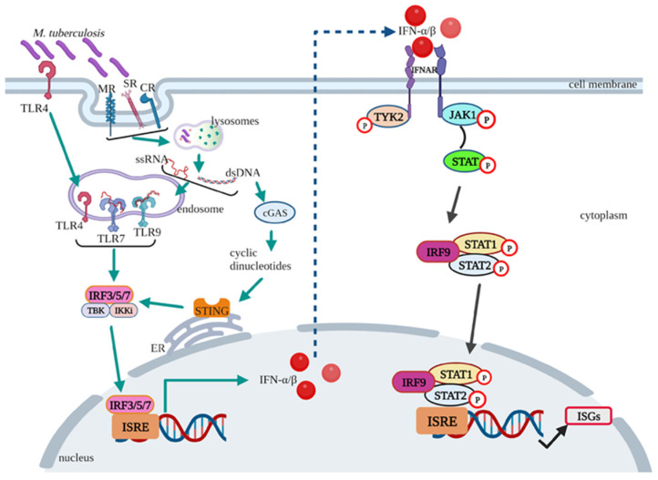
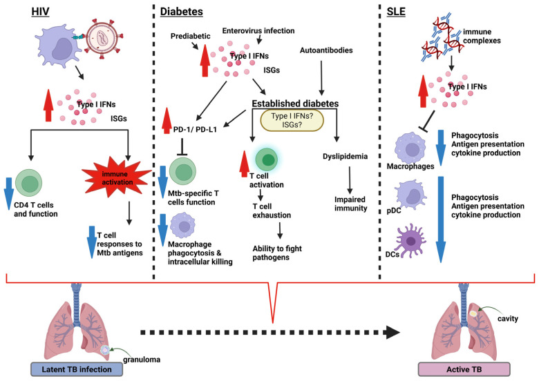
The identification of a type I interferon-induced transcriptomic signature in active tuberculosis suggests a potential role for these interferons in the pathogenesis of tuberculosis. Comorbidities such as human immunodeficiency virus, diabetes, systemic lupus erythematosus, end-stage renal disease, and coronavirus disease are epidemiologically linked to an increased risk for reactivation of latent tuberculosis infection. Notably, type I interferons are also implicated in the pathogenesis of these conditions, with a recognizable type I interferon transcriptomic signature. The mechanisms by which type I interferons in tuberculosis-risk-associated comorbidities may drive the progression of tuberculosis or maintenance of latent infection however remain largely unknown. This review summarizes the existing literature on the increased association between type I interferons, focusing on interferon-α and -β, and the heightened risk of tuberculosis reactivation. It also underscores the similarities in the immunopathogenesis of these comorbidities. A better understanding of these mechanisms is essential to guide the development of host-directed interferon therapies and improving diagnostic biomarkers in M. tuberculosis infection.

Keywords: COVID-19; HIV; diabetes; end-stage renal disease; interferon signature; silicosis; systemic lupus erythematosus; tuberculosis; type I interferon.


Conflict of interest statement

The authors declare no conflicts of interest.

Figures

Figure 1
Figure 1
Type I IFN induction and signalling pathway in M. tuberculosis infection. The figure illustrates the cascade of type I IFN production following M. tuberculosis infection and the subsequent induction of the IFN signalling pathway, resulting in the downstream production of ISGs. TLR—Toll-like receptor; MR—mannose receptor; SR—scavenger receptor; CR—complement receptor; ssRNA—single-stranded ribonucleic acid; dsDNA—double-stranded deoxyribonucleic acid; ER—endoplasmic reticulum; IFN—interferon, IFNAR—interferon-alpha receptor; IRF—interferon regulatory factor; ISRE—interferon-sensitive response element; TYK—tyrosine kinase; JAK—Janus kinase; STAT—signal transducer and activator of transcription; ISG—interferon-stimulated genes; cGAS—cyclic GMP-AMP synthase; STING—stimulator of IFN genes; TBK1—tyrosine kinase 1; IKK—IκB kinase, ISGs—interferon stimulated genes. Created with BioRender.com (Available from https://www.biorender.com/) (Accessed on 30 January 2025).
Figure 2
Figure 2
Summary of type I IFN responses in HIV, diabetes, and SLE with potential for reactivation of LTBI. The diagram illustrates the potential mechanisms by which type I IFN-associated diseases can lead to the reactivation of latent tuberculosis infection (LTBI) into active tuberculosis (TB). HIV interacts with pDCs and induces the production of increased levels of type I IFNs. These IFNs act by suppressing CD4 T cell counts and function or by causing immune activation, which suppresses M. tuberculosis-specific T cell responses. Diabetes: The production of type I IFNs is induced by either enterovirus infections associated with type 1 diabetes (T1D) or inflammation-mediated T2D. The IFNs act by increasing the PD-1/PD-L1 expression that impairs M. tuberculosis-specific function and macrophage phagocytosis and intracellular killing. IFNs also cause T cell exhaustion, impairing their ability to kill pathogens. SLE: Immune complexes and autoantibodies produced in SLE induce increased levels of type I IFNs. The IFNs reduce the capability of macrophages to carry out phagocytosis, antigen presentation, and cytokine production, and that of DCs and pDCs to migrate. The described mechanisms resulting from the type I IFNs in these diseases have the potential, in the presence of LTBI, to drive progression to active TB. Created with BioRender.com (Available from https://www.biorender.com/) (Accessed on 10 June 2025). IFN—interferon; ISGs—interferon stimulated genes; PD—programmed cell death protein; PD-L1—programmed death-ligand 1; DC—dendritic cell; pDC—plasmacytoid dendritic cell; Mtb—M. tuberculosis.

Similar articles

  • Systemic treatments for metastatic cutaneous melanoma.
    Pasquali S, Hadjinicolaou AV, Chiarion Sileni V, Rossi CR, Mocellin S. Pasquali S, et al. Cochrane Database Syst Rev. 2018 Feb 6;2(2):CD011123. doi: 10.1002/14651858.CD011123.pub2. Cochrane Database Syst Rev. 2018. PMID: 29405038 Free PMC article.
  • Type I and Type III Interferons Restrict SARS-CoV-2 Infection of Human Airway Epithelial Cultures.
    Vanderheiden A, Ralfs P, Chirkova T, Upadhyay AA, Zimmerman MG, Bedoya S, Aoued H, Tharp GM, Pellegrini KL, Manfredi C, Sorscher E, Mainou B, Lobby JL, Kohlmeier JE, Lowen AC, Shi PY, Menachery VD, Anderson LJ, Grakoui A, Bosinger SE, Suthar MS. Vanderheiden A, et al. J Virol. 2020 Sep 15;94(19):e00985-20. doi: 10.1128/JVI.00985-20. Print 2020 Sep 15. J Virol. 2020. PMID: 32699094 Free PMC article.
  • Measures implemented in the school setting to contain the COVID-19 pandemic.
    Krishnaratne S, Littlecott H, Sell K, Burns J, Rabe JE, Stratil JM, Litwin T, Kreutz C, Coenen M, Geffert K, Boger AH, Movsisyan A, Kratzer S, Klinger C, Wabnitz K, Strahwald B, Verboom B, Rehfuess E, Biallas RL, Jung-Sievers C, Voss S, Pfadenhauer LM. Krishnaratne S, et al. Cochrane Database Syst Rev. 2022 Jan 17;1(1):CD015029. doi: 10.1002/14651858.CD015029. Cochrane Database Syst Rev. 2022. Update in: Cochrane Database Syst Rev. 2024 May 2;5:CD015029. doi: 10.1002/14651858.CD015029.pub2. PMID: 35037252 Free PMC article. Updated.
  • Management of urinary stones by experts in stone disease (ESD 2025).
    Papatsoris A, Geavlete B, Radavoi GD, Alameedee M, Almusafer M, Ather MH, Budia A, Cumpanas AA, Kiremi MC, Dellis A, Elhowairis M, Galán-Llopis JA, Geavlete P, Guimerà Garcia J, Isern B, Jinga V, Lopez JM, Mainez JA, Mitsogiannis I, Mora Christian J, Moussa M, Multescu R, Oguz Acar Y, Petkova K, Piñero A, Popov E, Ramos Cebrian M, Rascu S, Siener R, Sountoulides P, Stamatelou K, Syed J, Trinchieri A. Papatsoris A, et al. Arch Ital Urol Androl. 2025 Jun 30;97(2):14085. doi: 10.4081/aiua.2025.14085. Epub 2025 Jun 30. Arch Ital Urol Androl. 2025. PMID: 40583613 Review.
  • NIH Consensus Statement on Management of Hepatitis C: 2002.
    [No authors listed] [No authors listed] NIH Consens State Sci Statements. 2002 Jun 10-12;19(3):1-46. NIH Consens State Sci Statements. 2002. PMID: 14768714

References

    1. The World Health Organization (WHO) World Health Organization Global Tuberculosis Report 2023. The World Health Organization (WHO); Geneva, Switzerland: 2023.
    1. The World Health Organization (WHO) World Health Organization Global Tuberculosis Report 2024. The World Health Organization (WHO); Geneva, Switzerland: 2024.
    1. World Health Organization . WHO: Operational Handbook on Tuberculosis. World Health Organization; Geneva, Switzerland: 2020.
    1. Migliori G.B., Ong C.W.M., Petrone L., D’ambrosio L., Centis R., Goletti D. The Definition of Tuberculosis Infection Based on the Spectrum of Tuberculosis Disease. Breathe. 2021;17:210079. doi: 10.1183/20734735.0079-2021. - DOI - PMC - PubMed
    1. O’Garra A., Redford P.S., McNab F.W., Bloom C.I., Wilkinson R.J., Berry M.P.R. The Immune Response in Tuberculosis. Annu. Rev. Immunol. 2013;31:475–527. doi: 10.1146/annurev-immunol-032712-095939. - DOI - PubMed

LinkOut - more resources