. 2025 Jul 23;20(7):e0328970.
doi: 10.1371/journal.pone.0328970. eCollection 2025.

A role for Myo-II zipper and spaghetti squash in Gliotactin-dependent Drosophila melanogaster wing hair planar cell polarity

Affiliations

A role for Myo-II zipper and spaghetti squash in Gliotactin-dependent Drosophila melanogaster wing hair planar cell polarity

Chase Holland et al. PLoS One. .

Abstract

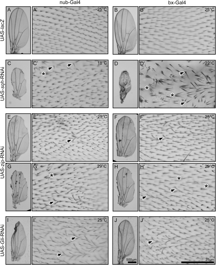
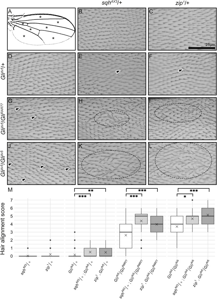
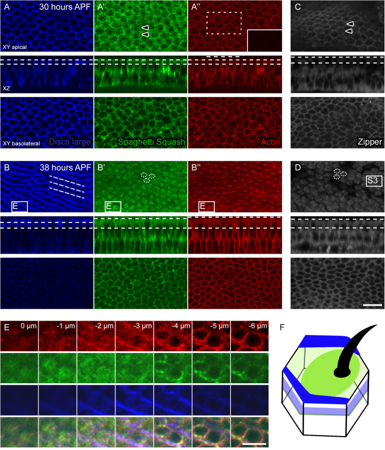
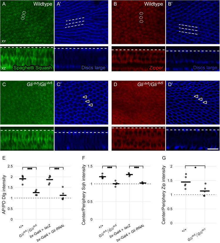
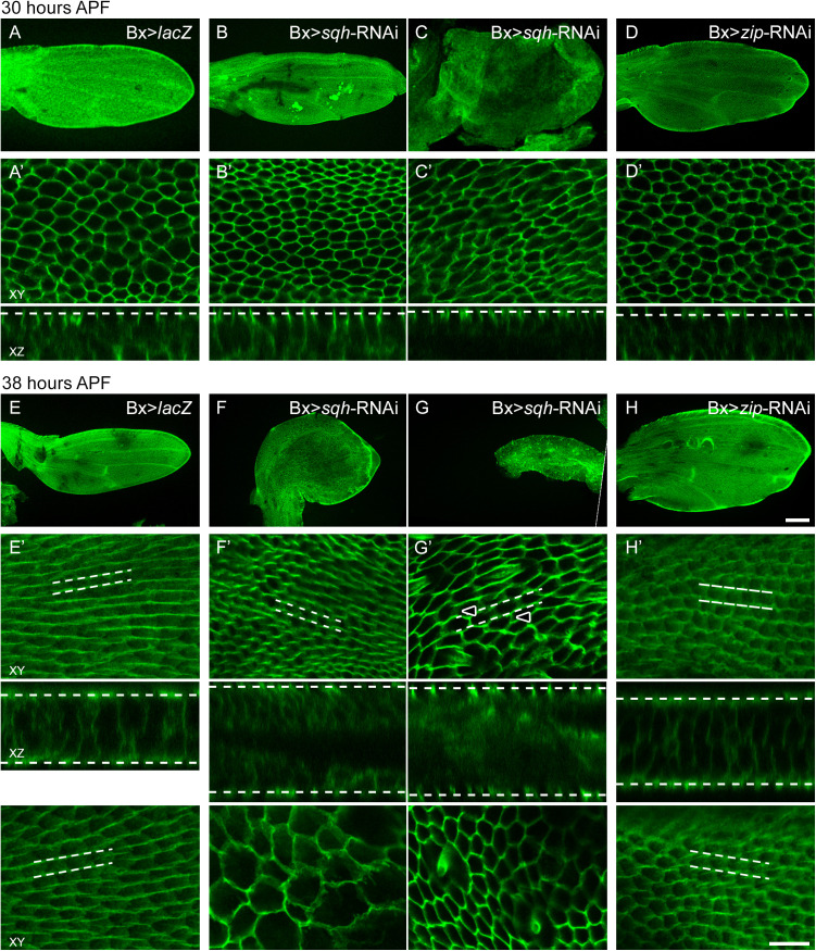
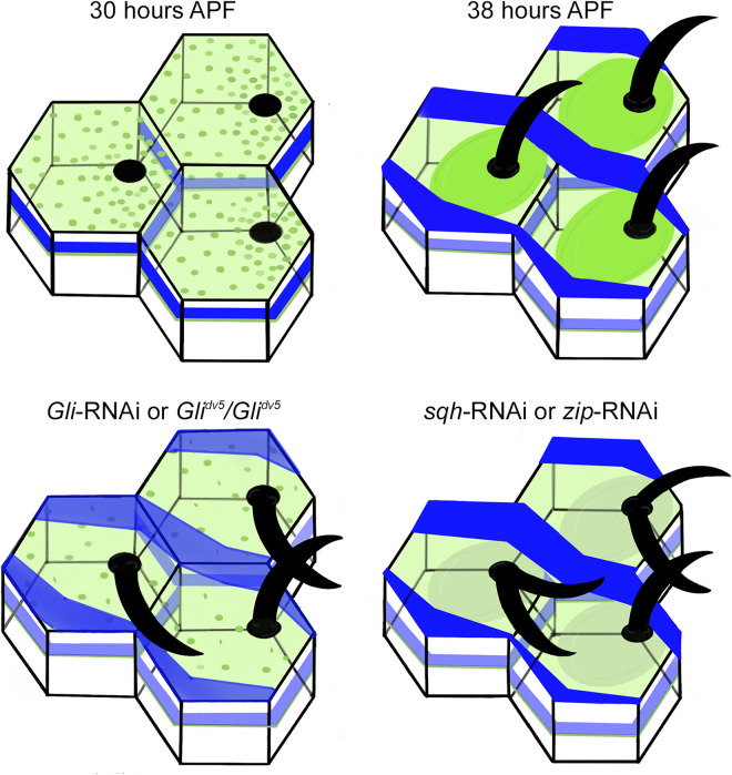
Planar cell polarity, polarization in the plane of an epithelium, is critical for tissue development. The Drosophila melanogaster wing epithelium is an important model system for planar cell polarity establishment and has greatly informed studies in vertebrates. The well-studied Frizzled-dependent and Fat - Dachsous - Four-jointed pathways establish proximal-to-distal polarity of wing hairs, while a Frizzled-independent mechanism mediated by septate junction proteins Gliotactin, Coracle, and Varicose is required for parallel alignment of neighboring hairs. In this study, we explore a requirement for the non-muscle myosin II proteins Spaghetti Squash and Zipper in wing hair planar cell polarity. We confirm a previously recognized role in hair initiation and demonstrate a second, novel Gliotactin-interacting requirement for spaghetti squash and zipper in parallel alignment. Immunolabeling experiments demonstrate that Spaghetti Squash and Zipper localize to the base of the developing hairs during the same time frame that septate junction proteins transiently relocalize to the apical cell surface. This localization is abrogated in Gliotactin loss-of-function genotypes. We propose that Gliotactin promotes Spaghetti Squash and Zipper accumulation at the cell apical surface during wing hair extension and that this apical Myosin-II complex stabilizes the developing hair base, maintaining parallel alignment of neighboring wing hairs.


Conflict of interest statement

The authors have declared that no competing interests exist.

Figures

Fig 1
Fig 1. Myo-II zip and sqh are required for multiple aspects of wing hair development.
Control (nub > lacZ or bx > lacZ) wings are flat and have smooth and consistent edges (A and B). Wing hairs have consistent proximal-to-distal directionality and neighboring hairs are aligned in parallel (A’ and B’). Wings with reduced Sqh or Zip or Gli were generated using wing-targeted RNAi. In contrast to wildtype wings, wings from flies with RNAi directed against either sqh or zip have an overall crumpled structure (e.g., D), serrated edges (e.g., C, E-H), and/or dark accumulations along the veins (e.g., F-H). RNAi wings also have patches of multiple wing hairs (asterisks in C’-H’) and wing hair misalignment (arrows in C’-H’). For both sqh and zip, the mutant phenotypes were more severe at higher temperatures (18°C, 22°C, 25°C, and 29°C were tested, with representative images shown). Wings from flies with RNAi directed against Gli were frequently crumpled, especially at the posterior distal wing edge (e.g., I). Patches of hairs had wing hair misalignment (see arrows in I’ and J’). Multiple wing hairs were not observed in Gli-RNAi wings. Calibration: 500 µm in A-J, 25 µm in A’-J’.
Fig 2
Fig 2. zip1 and sqhAX3 dominantly enhance Gli mutant wing hair misalignment.
(A) D. melanogaster wing veins separate the wing into seven regions (indicated by asterisks). (B-L) Wing hairs from heterozygotes for sqhAX3 (B), zip1 (C), and Glidv5 (D) have consistent proximal-to-distal directionality and parallel alignment of neighboring hairs. Flies doubly heterozygous for Glidv5 and either sqhAX3 or zip1 occasionally have neighboring hairs that tilt towards one another (E-F, arrows). Glidv5/GliRAR77 (G) and Glidv5/Glidv5 (J) mutant wings exhibit neighboring hairs that tilt towards one another. Gli mutants that additionally are heterozygous for either sqhAX3 (H, K) or zip1 (I, L) have increased hair misalignment, often in patches (dotted ellipses in H, I, K, L). B-L calibration: 25 µm. (M) Misalignment severity was quantified by counting the number of regions per wing with misaligned hairs. For box plots, center lines show the median values, box limits indicate the 25th and 75th percentiles, whiskers extend 1.5 times the interquartile range (IQR) from the 25th and 75th percentiles, and dots indicate outliers beyond the 1.5 IQR. X indicates the mean value. Kruskal-Wallis and Dunn’s tests were used to identify sqhAX3/+ and zip1/ + enhanced genotypes that significantly differed from the Gli mutant base (comparisons indicated by the brackets). * p = 0.017, ** p = 0.005, *** p < 0.001.
Fig 3
Fig 3. Sqh and Zip accumulate at the apical cell surface during pre-hair extension.
(A-D) Apical (XY apical), mid-cell (XY basolateral), and transverse (XZ) images of wildtype pupal wings at 30 and 38 hours APF. Dotted lines in the XZ image indicate the positions of the apical and basolateral XY planes. The inset in A” displays a plane immediately above the apical cell surface, at the position of the developing hairs (inset was imaged at the position indicated by the dashed rectangle). (E) Enlarged images of the boxed region in B-B” shown at descending 1 µm Z intervals, with the 0 µm position set at the level of the extended hairs above the apical cell surface. See S3A Fig for enlarged images of boxed region in D. (F) A cartoon summary of the Dlg (blue) and Sqh and Zip (green) localization at 38 hours APF. (A-F) In wildtype pupal wings at 30 hours APF, Actin (Phalloidin, red) and Dlg (blue) surround the cell periphery at both the apical and basolateral positions (A, A”). No extended hairs are present (A,” inset). Sqh-GFP (green, A’) and Zip-GFP (grey, C) surround the cell periphery, with diffuse patterns visible along the apical cell surface, with slight concentrations at the distal vertex (arrowheads). By 38 hours APF, the developing hairs (B,” E 0 µm) extend above the apical surface of each cell and Dlg (B, E −2 to −4 µm) forms ribbons below the apical surface of the wing (dotted parallel lines track three example ribbons). Unlike the diffuse pattern at 30 hours APF, Sqh-GFP (green, B’, E −1 µm) and Zip-GFP (grey, D) are present in circular accumulations near the cell surface (dotted ellipses outline example accumulations). Note that in A-A,” the apical planes were all imaged at the same Z position, while in B-B” the apical planes differed. This reflects a shift in the most apical position at which the relevant label was detected, which can be more clearly seen in E. At both 30 and 38 hours APF, Actin, Dlg, Sqh-GFP, and Zip-GFP are present around the cell periphery basolaterally (A-D bottom panels, E −5 to −6 µm). For both 30 and 38 hours, Actin, Dlg, and Sqh-GFP were imaged in a single wing, while Zip-GFP is shown in a different wing (also labeled with Dlg, as shown in S3A Fig). The bottom row in E indicates the Actin (red), Sqh-GFP (green), and Dlg (blue) merge. Calibration: 10 µm in A-D, 5 µm in E.
Fig 4
Fig 4. Gli is required for Sqh and Zip apical accumulation.
(A-D) Apical (XY) and transverse (XZ) sections of pupal wings at 38 hours APF. The dotted lines in the XZ images indicate the positions of the XY planes. In wildtype wings, Dlg (blue) accumulates in apical ribbons at 38 hours APF, with minimal to no Dlg present at proximodistal cell boundaries (A’, B’; dotted parallel lines track three example ribbons). Immediately apical to these ribbons, Sqh-GFP (green) and Zip-GFP (red) are present in circular accumulations (A and B; dotted ellipses outline example accumulations). In Glidv5/Glidv5, Dlg does not form continuous ribbons, but remains around the lateral cell surface (e.g., C’, D’). Arrowheads indicate residual accumulation at the proximodistal cell boundaries. Unlike in wildtype, Zip and Sqh do not accumulate apically in Glidv5/Glidv5, but remain diffuse at the apical surface (C, D). Calibration: 10 µm. (E-G) Quantification of Dlg, Sqh, and Zip apical localization. Dlg localization in proximal-distal ribbons was quantified as the ratio of fluorescence intensity along anteroposterior (AP) cell boundaries relative to proximodistal (PD) cell boundaries. Sqh-GFP and Zip-GFP localization in apical circular accumulations was quantified as a ratio of fluorescence intensity within a circular patch around the apical cell center (CC) relative to the apical cell periphery (CP). The intensity ratios were calculated for five wings per genotype (with five cells sampled and averaged per wing). For plots, dots indicate the average ratio per wing and horizontal lines show the mean values. * p = 0.038, *** p < 0.001 (Welch’s 2-sample t-test for Zip-GFP single pair analyses or ANOVA followed by a Tukey’s HSD test for Dlg and Sqh-GFP multiple pair analyses).
Fig 5
Fig 5. Inconsistent Dlg ribboning in wings with reduced sqh or zip.
Full wing (A-H) and apical and transverse sections (XY and XZ respectively, A’-H’) of Dlg-labeled pupal wings at 30 and 38 hours APF. The dotted lines in the XZ images indicate the positions of the XY planes. At 30 hours APF, control pupal wings have a consistent arrangement of hexagonal cells with Dlg surrounding the cell peripheries (A’). By 38 hours APF, Dlg is present in apical ribbons on both the dorsal and ventral wing surfaces (E’ top and bottom panels; dotted parallel lines track two example ribbons). At both 30 and 38 hours APF, the overall wing shape is flat and regularly patterned (A and E). In contrast, wings with RNAi directed against sqh have disruptions in overall wing shape (B, C, F, G) and cell size (e.g., compare cell size differences in the top and bottom panels of F’). Because of the variable phenotypes, two examples of bx > sqh-RNAi wings at each stage are shown. Wings with reduced zip had normal to slightly disrupted overall wing shape (D and H). Despite these wing shape distortions, in bx > sqh-RNAi and bx > zip-RNAi wings Dlg was normally localized to the cell periphery at 30 hours APF (B’, C’). By 38 hours APF, localization patterns were varied in bx > sqh-RNAi wings. In some wing layers – particularly those with severe cell distortion – Dlg remained along the entire cell periphery (e.g., bottom panels in F’, G’). In other wings, apical ribboning was evident, either fully (e.g., top panel in F’) or partially (e.g., top panel in G’; arrowheads indicate examples of lower intensity of Dlg signal at proximodistal cell boundaries). In bx > zip-RNAi wings at 38 hours APF (H’), Dlg apical ribboning was present, but less continuous than in controls. In E’-H’, the top and bottom XY panels are apical sections of the dorsal and ventral wing surfaces taken at the same XY position. Because the Dlg localization pattern was identical in both surfaces, only a single wing surface is shown in A’-D’. Calibration: 100 µm in full wing panels, 10 µm in all others.
Fig 6
Fig 6. A summary model of Myo-II in wing hair development.
At 30 hours APF, when hair development initiates, Dlg (blue) is restricted to the cell periphery and Sqh and Zip (green) are present around the cell periphery and diffusely at the apical cell surface. By 38 hours APF, during hair extension, Dlg localizes in apical proximal-distal ribbons, with lower levels persisting around the cell periphery. Zip and Sqh accumulate at the apical cell surface in circular disks around the base of the developing hairs. In wings with reduced Gli, Dlg ribbons are weakly present or absent, Sqh and Zip are diffuse at the apical cell surface, and wing hairs do not maintain parallel alignment. In wings with reduced sqh or zip that maintain normal cell morphology, Dlg localizes in apical ribbons, but wing hairs exhibit both multiple wing hair and hair misalignment phenotypes.

Similar articles

References

    1. Butler MT, Wallingford JB. Planar cell polarity in development and disease. Nat Rev Mol Cell Biol. 2017;18(6):375–88. doi: 10.1038/nrm.2017.11 - DOI - PMC - PubMed
    1. Derish I, Lee JKH, Wong-King-Cheong M, Babayeva S, Caplan J, Leung V, et al. Differential role of planar cell polarity gene Vangl2 in embryonic and adult mammalian kidneys. PLoS One. 2020;15(3):e0230586. doi: 10.1371/journal.pone.0230586 - DOI - PMC - PubMed
    1. Devenport D. Tissue morphodynamics: Translating planar polarity cues into polarized cell behaviors. Semin Cell Dev Biol. 2016;55:99–110. doi: 10.1016/j.semcdb.2016.03.012 - DOI - PMC - PubMed
    1. Hirota Y, Meunier A, Huang S, Shimozawa T, Yamada O, Kida YS. Planar polarity of multiciliated ependymal cells involves the anterior migration of basal bodies regulated by non-muscle myosin II. Development. 2010;137(18):3037–46. - PubMed
    1. Kunimoto K, Bayly RD, Vladar EK, Vonderfecht T, Gallagher A-R, Axelrod JD. Disruption of Core Planar Cell Polarity Signaling Regulates Renal Tubule Morphogenesis but Is Not Cystogenic. Curr Biol. 2017;27(20):3120-3131.e4. doi: 10.1016/j.cub.2017.09.011 - DOI - PMC - PubMed

MeSH terms

LinkOut - more resources