. 2025 Jul 23;21(7):e1013219.
doi: 10.1371/journal.ppat.1013219. eCollection 2025 Jul.

A functional interleukin-4 homolog is encoded in the genome of infectious laryngotracheitis virus: Unveiling a novel virulence factor

Affiliations

A functional interleukin-4 homolog is encoded in the genome of infectious laryngotracheitis virus: Unveiling a novel virulence factor

Jeremy D Volkening et al. PLoS Pathog. .

Abstract

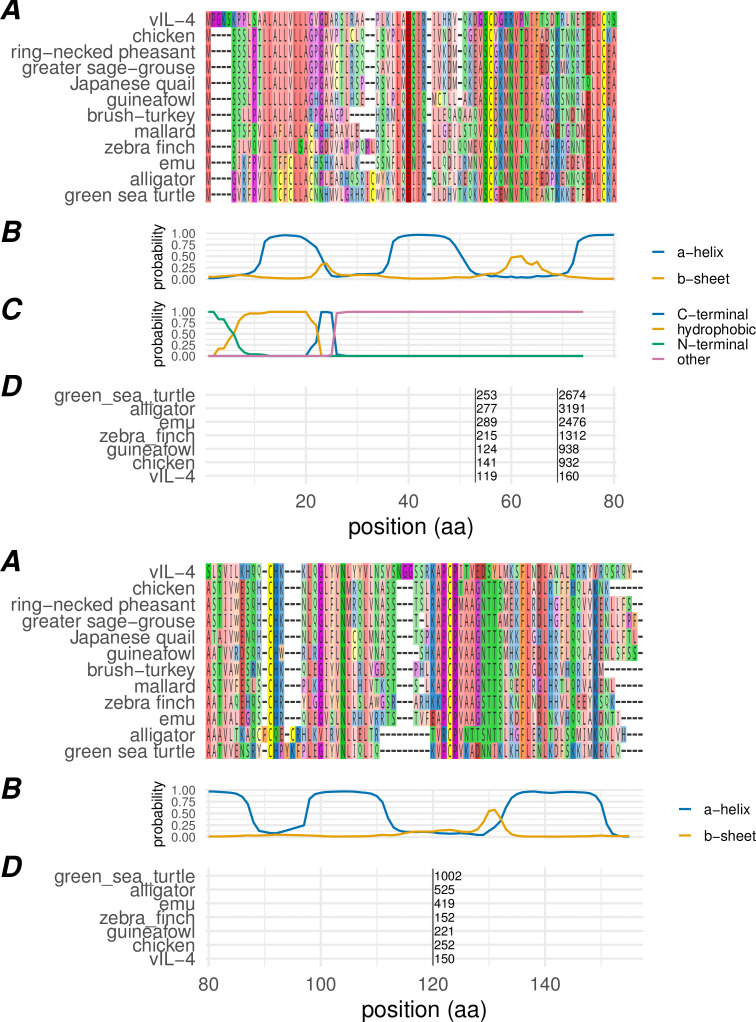
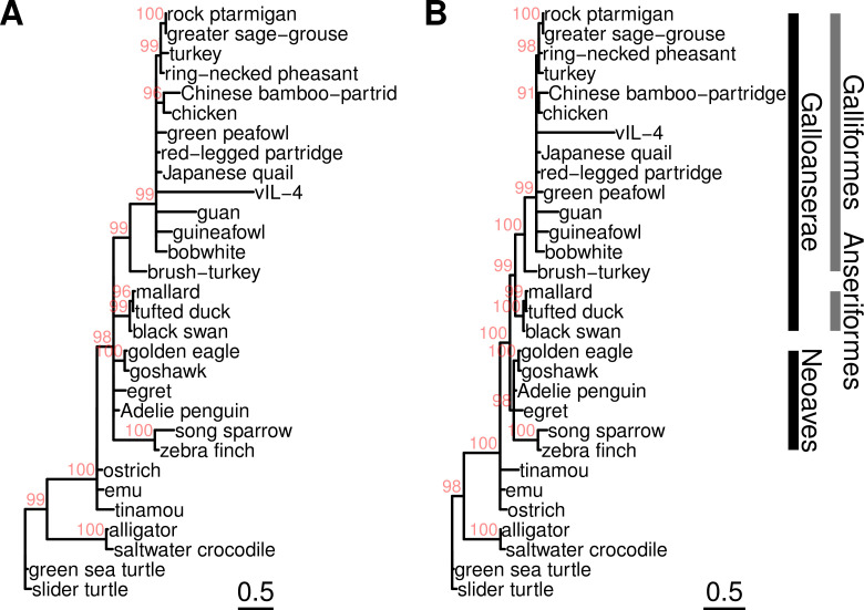
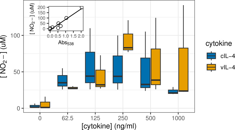
Herpesviruses have evolved numerous immune evasion tactics, persisting within their hosts through self-perpetuating strategies. One such tactic involves acquiring functional copies of host genes encoding cytokines such as IL-6 (HHV-8), IL-10 (HHV-4, HHV-5), and IL-17 (SaHV-2). These viral mimics, or virokines, can bind to cellular receptors, modulating the natural cytokine signaling to manipulate the immune response in favor of the virus or stimulate target cell growth to enhance virus replication. In the course of full-length cDNA sequencing of infectious laryngotracheitis virus (ILTV) transcripts, a previously unknown highly spliced gene was discovered in the viral genome predicted to encode a 147 amino acid protein with similarity to vertebrate interleukin-4. The three-intron gene structure was precisely conserved with chicken and other vertebrate IL-4 homologs, and the amino acid sequence displayed structural conservation with vertebrate homologs at the primary, secondary, and tertiary levels based on computational modeling. The viral IL-4 gene was subsequently identified in all sequenced ILTV genomes. The mature transcript was highly expressed both in vitro and in vivo, and protein expression in infected cells was confirmed using LC-MS/MS. Phylogenetic analyses, along with the conserved gene structure, suggested direct capture from a Galliformes host. Functionally, an LPS-stimulation assay showed that the expressed viral IL-4 homolog stimulated nitric oxide production in a macrophage cell line at comparable levels to recombinant chicken IL-4. A recombinant virus lacking vIL-4 exhibited slightly higher titers in cell culture compared to the parental strain. In vivo bird studies demonstrated reduced pathogenicity of the vIL-4 knockout compared to wildtype. These results represent the first report of a previously unknown virokine encoded in the ILTV genome expressing a functional IL-4 homolog and virulence factor.


Conflict of interest statement

The authors have declared that no competing interests exist.

Figures

Fig 1
Fig 1. Alignment of experimental IsoSeq transcripts and MS/MS peptides to the ILTV UL/IRS genomic junction.
Tracks shown are (from top to bottom) genomic coordinates; genomic regions; existing annotations; 1874C5 IsoSeq models; CEO (Laryngo-Vac) IsoSeq models; LC-MS/MS experimentally identified peptides; published LJS09 RNA-Seq read depth. IsoSeq models are colored by strand (blue: + , red: -) and shaded by supporting read count (darker = higher count). The primary 4-exon vIL-4 transcript (center, forward strand) is roughly 100x more abundant than the alternative isoform models in 1874C5. In CEO (Laryngo-Vac), where there was lower viral abundance overall, only the dominant isoform was detected. Most of the theoretically detectable tryptic peptides from vIL-4 were detected at 1% FDR using bottom-up LC-MS/MS (purple features). Also shown (bottom track) is an alignment of previously published short-read RNA-Seq data from strain LJS09 demonstrating expression of the vIL-4 transcript in an additional strain. Only a single replicate from this experiment is shown for the sake of brevity, but all four infected replicates showed similar coverage.
Fig 2
Fig 2. Highly expressed in vivo viral transcripts.
Shown are the 8 most highly expressed ILTV transcripts by median transcripts per million transcripts (TPM) as a distribution of (A) viral TPM and (B) ordinal rank of TPM. RNA-Seq expression data is from ocular inoculation of birds with 1874C5 followed by sampling at 3, 5, and 7 days post-infection (d.p.i.). In this experiment, vIL-4 is the fifth most abundant transcript expressed, following two origin-associated transcripts, glycoprotein G, and VP22.
Fig 3
Fig 3. Primary, secondary, and genomic structure of the viral interleukin-4 homolog.
(A) Amino acid alignment of vIL-4 with homologs from selected species. (B) Secondary structure probability of the vIL-4 amino acid sequence as predicted by JPRED2. (C) Signal peptide domain probability as predicted by SignalP 6.0; indicated are the N-terminal domain [‘N’], hydrophobic domain [‘H’], and C-terminal domain [‘C’]. (D) Location of genomic introns in a representative selection of homologs, translated into amino acid space. Black vertical bars indicate the intron position, with the intron length indicated to the right of each bar. Intron locations within the amino acid alignment were identical for all homologs examined, including those not shown.
Fig 4
Fig 4. Comparative predicted tertiary structures.
(A) Empirical crystal structure of human IL-4 (PDB:2B8U); cysteines/disulfide bonds shaded yellow. (B) AlphaFold model of vIL-4 shaded by pLDDT from low (red) to high (blue); cysteines/disulfide bonds shaded yellow. ILTV vIL-4 is predicted to share the same four-alpha-helix, anti-parallel beta sheet arrangement as the empirically modeled hIL-4. Like all avian IL-4 homologs, it lacks the cysteines at both protein termini that form a third disulfide bond in human IL-4 (panel A lower-left). (C) Superimposed predicted structures of vIL-4 (blue) and chicken IL-4 (green) in complex with chicken IL-4Rα (transparent purple) and γc (transparent red) receptor subunits (Type I receptor complex), as predicted by AlphaFold and aligned by the RCSB Pairwise Structure Alignment tool (jFATCAT/rigid). Disulfide bridges are shown in yellow.
Fig 5
Fig 5. ML trees of vIL-4 and interleukin-4 homologs from a selection of representative bird and reptilian genomes.
(A) From amino acid alignment, built with IQTREE using the ‘Q.bird+I+G4’ model as selected by ModelFinder, ultrafast bootstrapping (1000 iterations). (B) From coding nucleotide alignment, built with IQTREE using the ‘GTR+I+R’ model as selected by ModelFinder, ultrafast bootstrapping (1000 iterations). Branches with UFBoot support (in red) < 90 were collapsed.
Fig 6
Fig 6. ML tree of amino acid sequences of vIL-4 in sequenced ILTV genomes.
An intact gene structure and inframe coding sequence was found in all 89 complete ILTV genomes examined (after removing known recombinants, duplicates, and the original published mosaic genome). Most of the amino acid sequences were identical and are shown as a single ‘Serva-like’ cluster. Only a handful of variants characterized the remaining sequences. Each branchpoint on the tree represents one or several amino acid variants, labeled in blue. The tree was built with IQTREE using the ‘Q.bird’ model as selected by ModelFinder, ultrafast bootstrapping (10000 iterations). Support values shown in gray are UFBoot estimations.
Fig 7
Fig 7. Comparison of vIL-4, cIL-4 and cIL-13 amino acid similarity and predicted receptor interactions.
Panels A-E are aligned on amino acid position. (A) Secondary structure probability of the vIL-4 amino acid sequence as predicted by JPRED2. (B) Positions of identical amino acids between indicated aligned sequence pairs. (C) Positions of predicted atomic interactions between indicated ligand and IL-4 type I receptor subunit pairs (based on AlphaFold multimeric structural predictions). (D) Positions of predicted atomic interactions between indicated ligand and IL-4 type II receptor subunit pairs (based on AlphaFold multimeric structural predictions). (E) Amino acid alignment of vIL-4, cIL-4, and cIL-13 without signal peptides. (F) Number of predicted hydrogen bonds (first value) and salt bridges (second value) between two cytokine/virokine ligands and each type I receptor subunit. (G) Number of predicted hydrogen bonds (first value) and salt bridges (second value) between three cytokine/virokine ligands and each type II receptor subunit. (H) Calculated fraction identity and similarity (see methods for calculation) between each pairwise set of cytokine/virokine sequences.
Fig 8
Fig 8. Stimulation by IL-4 of nitric oxide production in macrophages.
The Griess assay was used to measure NO2- as an indicator of NO production at varying concentrations of either chicken IL-4 (cIL-4) or viral IL-4 (vIL-4) and in the presence of 2.5 µg/mL LPS. Inset: standard curve of Griess reagent used to calculate NO2- concentrations (R2 = 0.92).
Fig 9
Fig 9. Growth kinetics of 1874C5 parental and ΔvIL-4 ILTV strains in the LMH cell line.
LMH cells (1.4x 10⁶ cells per well) were inoculated in triplicate with 1874C5 parental strain and ΔvIL-4 strain at MOI of 0.002. After absorption for 1 hour at 39°C and 5% CO2, cells were washed and incubated for 0, 24, 48, 72 and 96 hours. At each time point supernatant and cells were collected (n = 3) and (A) viral titer and (B) viral genome copy number were determined for each strain at each time point. Individual replicates and corresponding nonlinear fit curves are shown.
Fig 10
Fig 10. Pathogenicity of 1874C5 (parent) and vIL-4 gene-deleted strain (Δ vIL-4) in specific pathogen free (SPF) chickens after intratracheal inoculation at 24 days of age.
(A) Percent survival for chickens inoculated with the ΔvIL-4 strain (n = 17) was 47.1% and for chickens inoculated with the parent strain (n = 15) was 33.3%. (B) Average clinical sign scores (CSS) per group of chickens from 2 to 7 days post-intratracheal inoculation. Although not significant, average CSS induced by the 1874C5 strain were higher than average scores induced by the knockout. (C) Viral genome load in trachea three days post-intratracheal inoculation. (D) Viral genome load in trachea five days postintratracheal inoculation. (E) Comparison of parent 1874C5 genome load at 3- and 5-days post-inoculation. (F) Comparison of ΔvIL-4 genome load at 3- and 5-days post-inoculation. Individual replicate loads indicated by points; column heights indicate the average trachea viral genome load per group. No significant (ns) differences (p > 0.05) were detected between viral genome load of the 1874C5 parent and the ΔvIL-4 mutant at either 3- or 5-days post-inoculation. Significant differences (p < 0.05) were detected for the average viral genome load of ΔvIL-4 in the trachea at 3- vs 5-days post-inoculation.
Fig 11
Fig 11. Histopathological lesions in the trachea induced by parental 1874C5 and ΔvIL-4 at four days post intratracheal inoculation.
Photomicrographs are H&E-stained upper trachea sections from chickens infected with parental 1874C5 (A & B) or ΔvIL-4 (C & D) at 20x magnification with image scale bar of 100 µm. (C) inset: Inserted photomicrograph at 40x magnification with image scale bar of 50 µm; underscores the presence of mucosal epithelium and goblet cells with few syncytia and intranuclear inclusion bodies still attached distal of the submucosa.

Similar articles

References

    1. Garcia M, Spatz S, Guy JS. Infectious laryngotracheitis. In: Swayne DE, Glisson JR, McDougald LR, Nolan LK, Suarez DL, Nair V, editors. Diseases of poultry. Ames, IA: Wiley-Blackwell; 2013.
    1. Fahey KJ, Bagust TJ, York JJ. Laryngotracheitis herpesvirus infection in the chicken: the role of humoral antibody in immunity to a graded challenge infection. Avian Pathol. 1983;12(4):505–14. doi: 10.1080/03079458308436195 - DOI - PubMed
    1. Bagust TJ, Jones RC, Guy JS. Avian infectious laryngotracheitis. Rev Sci Tech. 2000;19(2):483–92. doi: 10.20506/rst.19.2.1229 - DOI - PubMed
    1. Honda T, Okamura H, Taneno A, Yamada S, Takahashi E. The role of cell-mediated immunity in chickens inoculated with the cell-associated vaccine of attenuated infectious laryngotracheitis virus. J Vet Med Sci. 1994;56(6):1051–5. doi: 10.1292/jvms.56.1051 - DOI - PubMed
    1. Fahey KJ, York JJ. The role of mucosal antibody in immunity to infectious laryngotracheitis virus in chickens. J Gen Virol. 1990;71 ( Pt 10):2401–5. doi: 10.1099/0022-1317-71-10-2401 - DOI - PubMed

LinkOut - more resources