. 2025 Jul 21;301(9):110511.
doi: 10.1016/j.jbc.2025.110511. Online ahead of print.

N4BP1 is a nucleocytoplasmic shuttling protein and recognizes aggregates of the ubiquitin-like protein NEDD8 to protect cells under heat shock

Affiliations

N4BP1 is a nucleocytoplasmic shuttling protein and recognizes aggregates of the ubiquitin-like protein NEDD8 to protect cells under heat shock

Xiaohong Guo et al. J Biol Chem. .

Abstract

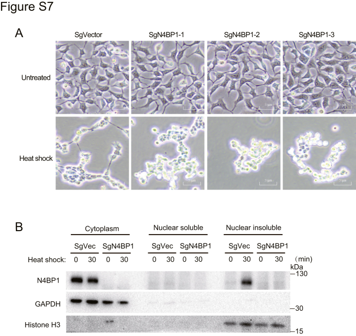
N4BP1 (NEDD4-binding partner 1) is a key checkpoint for proper inflammatory responses; however, its cellular localization, biologic nature, and functions in other cellular processes remain largely unknown. In this study, we demonstrate that N4BP1 is a nucleocytoplasmic shuttling protein. Treatment with leptomycin B induces nuclear accumulation of N4BP1, and the region responsible for its nuclear distribution maps to amino acids 151 to 338. Further analysis identified a nuclear localization signal (NLS) spanning residues 279 to 299. Deletion or mutation of this NLS abolishes N4BP1 nuclear import, while fusing the NLS to GFP is sufficient to drive GFP into the nucleus. Notably, we found that N4BP1 forms protein aggregates in both the cytoplasm and nucleus. These aggregates lack ubiquitin-modified proteins but instead colocalize with NEDD8-modified proteins. Consistently, N4BP1 aggregates contain cullin-1 and cullin-2. The CoCUN domain is essential for recognizing neddylated proteins and mediating N4BP1 aggregate formation. N4BP1 aggregates exhibit liquid-liquid phase separation, as evidenced by their sensitivity to 1,6-hexanediol (an liquid-liquid phase separation inhibitor). Smaller N4BP1 aggregates can fuse into larger one and reassemble after 1,6-hexanediol-induced disruption. Furthermore, heat shock promotes N4BP1 aggregation, which confers cellular protection under stress conditions. Taken together, our findings reveal that N4BP1 is a nucleocytoplasmic shuttling protein. N4BP1 forms protein aggregates that contain neddylated proteins such as cullin-1 and cullin-2. This study uncovers the previously unrecognized role of N4BP1 in organizing neddylated protein aggregates and highlights its functional significance in stress adaption.

Keywords: N4BP1; NEDD8; aggregates; heat shock; nucleocytoplasmic shuttling.


Conflict of interest statement

Conflict of interest The authors declare that they have no conflict of interest with the contents of this article.

Figures

Figure 1
Figure 1
N4BP1 shuttles between cytoplasma and nuclear insoluble fraction.A, 293T and HeLa cells were transfected with plasmids encoding FLAG-N4BP1, and the distribution of N4BP1 was determined by immunofluorescence using an anti-FLAG antibody. The images were acquired using a fluorescence microscope. Scale bar is 10 μm. B, the distribution of FLAG-N4BP1 in control and stably expressed 293T cells were determined by immunofluorescence using an anti-FLAG antibody. The images were acquired using a confocal microscope. Scale bar is 10 μm. C, 293T and HeLa cells were transfected with plasmids encoding FLAG-N4BP1, followed by treatment with 10 μM leptomycin B, importazole, or ivermectin for 1 h. The distribution of N4BP1 was determined by immunofluorescence using an anti-FLAG antibody. The images were acquired using a fluorescence microscope. Scale bar is 10 μm. D, quantification of N4BP1 nuclear-only cells in (C). N4BP1 nuclear-only cells were counted from 30 cells. Top: The radio of N4BP1 nuclear-only cells in FLAG-N4BP1-expressing 293T cells before and after drug treatment. Bottom: The radio of N4BP1 nuclear-only cells in FLAG-N4BP1-expressing HeLa cells before and after drug treatment. ∗∗∗∗p < 0.0001. E, 293T cells were transfected with plasmids encoding FLAG-N4BP1. The distribution of N4BP1 was determined by WB after biochemical fractionation of cytoplasm and nuclear extracts. F, HeLa cells were transfected with plasmids encoding FLAG-N4BP1 and then treated with 10 μM leptomycin B for 24 h. The distribution of N4BP1 was determined by WB after nuclear and cytoplasmic fractionation and insoluble component extraction. G, HeLa cells were treated with 10 μM leptomycin B for 24 h. The distribution of N4BP1 was determined by WB after nuclear and cytoplasm fractionation and insoluble component extraction. Data in (A and E) are representative of four independent experiments. Data in (B and C, F and G) are representative of three independent experiments. N4BP1, NEDD4-binding partner 1.
Figure 2
Figure 2
N4BP1 nuclear import relies on a domain between amino acids 151 to338.A, schematic representations of full-length N4BP1, N4BP1 deletion mutants, and N4BP1 point mutants. B, HeLa cells were transfected with plasmids encoding FLAG-tagged full-length N4BP1 or its deletion mutants. N4BP1 localization was determined by immunofluorescence using an anti-FLAG antibody. The images were acquired using a fluorescence microscope. Scale bar is 10 μm. C, HeLa cells were transfected with plasmids encoding FLAG-N4BP1, KH-only, or KH-UBA were treated with 10 μM leptomycin B for 4 h. N4BP1 localization was acquired using immunofluorescence (anti-FLAG). Images were captured with a fluorescence microscope. Scale bar is 10 μm. D, quantification of N4BP1 nuclear-exclusive localization in (C). Thirty FLAG-N4BP1-positive HeLa cells were counted. The ratio of cells displaying nuclear-only N4BP1 before and after leptomycin B treatment is shown. ∗∗p < 0.01; ∗∗∗∗p < 0.0001. E, HeLa cells were transfected with FL-N4BP1, N4BP1-L350A, or N4BP1-L379/380A and then treated with 10 μM leptomycin B for 4 h. N4BP1 distribution was assessed by immunofluorescence (anti-FLAG). Images were obtained using a confocal microscope. Scale bar is 10 μm. Data in (B, C, and E) are representative of three independent experiments. N4BP1, NEDD4-binding partner 1.
Figure 3
Figure 3
The NLS (279-299aa) of N4BP1 is important for its nuclear distribution.A, schematic diagrams of full-length N4BP1 and its NLS-deleted mutant (dNLS; residues 279-299: ERICQKRRFSDSEERHTKKQF). B, schematics of wildtype N4BP1 and NLS region mutants (Mut1-3). C, HeLa cells transfected with FL-N4BP1, dNLS, Mut1, Mut2, or Mut3 were analyzed by immunofluorescence (anti-FLAG). Images were observed by a confocal microscope. Scale bar is 10 μm; D, quantification of nuclear-exclusive N4BP1 in (C). Thirty FLAG-N4BP1-positive HeLa cells were counted. The ratio of nuclear-only cells posttransfection is shown. ∗∗∗∗p < 0.0001. E, 293T cells transfected with FL-N4BP1, dNLS, Mut1, Mut2, or Mut3 were analyzed by immunofluorescence (anti-FLAG). Images were observed by a confocal microscope. Scale bar is 10 μm; F, quantification of nuclear-exclusive N4BP1 in E. Thirty FLAG-N4BP1-positive 293T cells were counted. ∗∗∗∗p < 0.0001. G, HeLa cells transfected with GFP or GFP-NLS (279-299) were imaged by a fluorescence microscope. Scale bar is 10 μm. H, quantification of nuclear-exclusive GFP-NLS localization in (G). Thirty GFP-positive cells were counted. ∗∗∗∗p < 0.0001. Data in (C, E, and G) are representative of three independent experiments. N4BP1, NEDD4-binding partner 1; NLS, nuclear localization signal.
Figure 4
Figure 4
N4BP1 can form aggregates and N4BP1 distribution is excluded with DNA at nucleus.A, 293T and HeLa cells were transfected with plasmids encoding FLAG-N4BP1 and the aggregates of N4BP1 were determined by immunofluorescence (anti-FLAG). The images were observed by a confocal microscope. Scale bar is 10 μm. B, HeLa cells were transfected with plasmids encoding FLAG-N4BP1 and then treated with 3% of 1,6-hexanediol for 0, 5, 10, or 20 min. The aggregates of N4BP1 were determined by immunofluorescence (anti-FLAG). The images were observed by a confocal microscope. Scale bar is 10 μm; C, quantification of aggregate-positive cells in (B). Fifty FLAG-N4BP1-positive cells were counted. The ratio after 1,6-HD treatment is shown. ∗∗∗∗p < 0.0001. D, time-lapse imaging of N4BP1-GFP aggregate dissolution and recovery in 293T cells treated with 5% 1,6-HD. Scale bar is 10 μm. E, time-lapse imaging of N4BP1-GFP aggregates in 293T cells. Scale bar is 10 μm (top) or 2 μm (bottom). F, HeLa cells were transfected with plasmids encoding FLAG-N4BP1 and then treated with 3% of 1,6-hexanediol for 0, 10, or 30 min. The distribution of N4BP1 was determined by WB after nuclear and cytoplasmic fractionation assays and insoluble component extraction. G, HeLa cells were transfected with plasmids encoding FLAG-N4BP1, and the aggregates of N4BP1 was determined at nucleus by immunofluorescence using antibody against FLAG. The images were observed by a confocal microscope. Scale bar is 3 μm (top) or 1 μm (bottom). H, relative fluorescence intensity at the white dotted line (G) was quantified by using Image J. Data in (A) are representative of five independent experiments. Data in (B, DG) are representative of three independent experiments. N4BP1, NEDD4-binding partner 1; 1,6-HD, 1,6-hexanediol.
Figure 5
Figure 5
N4BP1 recognizes NEDD8-modifiedproteins to form aggregates.A, HeLa cells were transfected with plasmids encoding FLAG-N4BP1. Top: The immunofluorescence of NEDD8 (green) costained with FLAG-N4BP1 (red); Middle: The immunofluorescence of FLAG-N4BP1 (green) costained with Ub (red); Bottom: The immunofluorescence of NEDD8 (green) costained with Ub (red). The images were observed by a confocal microscope. Scale bar is 8 μm. B, HeLa cells expressing FLAG-N4BP1 were treated with 2 μM MLN4924 for 24 h. The immunofluorescence of NEDD8 (green) costained with FLAG-N4BP1 (red), and the images were observed by a confocal microscope. Scale bar is 10 μm. C, quantification of aggregate-positive cells in (B). Twenty FLAG-N4BP1-positive cells were counted. ∗∗p < 0.01. D, HeLa cells expressing FLAG-N4BP1 were treated with 3% of 1,6-hexanediol for 0, 10, or 30 min. The immunofluorescence of NEDD8 (green) costained with FLAG-N4BP1 (red), and the images were observed by a confocal microscope. Scale bar is 10 μm. E, quantification of aggregate-positive cells in (D). Twenty cells were counted. ∗∗∗p < 0.001 F, HeLa cells were transfected with plasmids encoding FLAG-N4BP1. The immunofluorescence of CUL1 and CUL2 (green) costained with FLAG-N4BP1 (red), and the images were observed by a confocal microscope. Scale bar is 10 μm. Data in (A) are representative of five independent experiments. Data in (B, D, and F) are representative of three independent experiments. N4BP1, NEDD4-binding partner 1.
Figure 6
Figure 6
The P866 of CoCUN domain is required for N4BP1 to recognized NEDD8.A, HeLa cells were transfected with plasmids encoding FLAG-N4BP1 or different N4BP1 deletion mutants. The immunofluorescence of NEDD8 (green) costained with FLAG-N4BP1 (red), and the images were observed by a confocal microscope. Scale bar is 8 μm. B, 293T cells were transfected with plasmids encoding FLAG-N4BP1 or NYN-CoCUN. The distribution of N4BP1 and NEDD8 were determined by WB after nuclear and cytoplasmic fractionation assays and insoluble component extraction. C, schematic representations of N4BP1 point mutants in KH, UBA, NYN, and CoCUN domains. D, HeLa cells were transfected with plasmids encoding FLAG-N4BP1-FL, G71D, G93D, or P866A. The immunofluorescence of NEDD8 (green) costained with FLAG-N4BP1 (red), and the images were observed by a confocal microscope. Scale bar is 8 μm. E, HeLa cells were transfected with plasmids encoding FLAG-N4BP1 or NYN-CoCUN. The immunofluorescence of CUL1 (green) costained with FLAG-N4BP1 (red), and the images were observed by a confocal microscope. Scale bar is 10 μm. F, HeLa cells were transfected with plasmids encoding FLAG-N4BP1 or NYN-CoCUN. The immunofluorescence of CUL2 (green) costained with FLAG-N4BP1 (red), and the images were observed by a confocal microscope. Scale bar is 10 μm. Data in (A, DF) are representative of three independent experiments. Data in (B) are representative of two independent experiments. N4BP1, NEDD4-binding partner 1.
Figure 7
Figure 7
N4BP1/NEDD8 aggregates play important role in heat shock.A, control and N4BP1 stably knocking down HaCaT cells were heated at 42 °C, and cell death was monitored hourly for 3 h by bright-field microscopy. Scale bar is 50 μm. B, control and N4BP1 stably knocking down HaCaT cells were treated with 2 μM MLN4924 for 6 h and then were treated in 42 °C. Cell death was monitored hourly for 3 h by bright-field microscopy. Scale bar is 50 μm. C, control and N4BP1-overexpression 293T cells were heated at 42 °C for 0.5 h, and cell death was observed by an optical microscope. Scale bar is 50 μm. D, 293T cells were transfected with plasmids encoding FLAG-N4BP1 and then heated at 42 °C for 20 min. Cell death was determined by immunofluorescence (anti-FLAG). The images were observed by a fluorescence microscope. Scale bar is 50 μm. E, quantification analysis of FLAG-N4BP1-positive cells in (D). One hundreds of cells from three different fields were counted. The ratio of FLAG-N4BP1-positive cells before and after heat shock were presented. F, 293T cells were treated or untreated in 42 °C for 30 min. The distribution of N4BP1 was determined by WB after nuclear and cytoplasmic fractionation assays and insoluble component extraction. G, control and N4BP1 stably knocking down HaCaT cells were heated at 42 °C for 2 h, and the aggregates of NEDD8 were determined by immunofluorescence (anti-NEDD8). The images were observed by a confocal microscope. Scale bar is 4 μm. H, 293T cells were transfected with plasmids encoding FLAG-N4BP1 or NYN-CoCUN and then heated at 42 °C for 0.5 h. Cell death was observed by an optical microscope. Scale bar is 50 μm. I, 293T cells were transfected with plasmids encoding FLAG-N4BP1 or P866A mutant and then heated at 42 °C for 20 min. Cell death was observed by an optical microscope. Scale bar is 50 μm. Data in (AD, FI) are representative of three independent experiments. N4BP1, NEDD4-binding partner 1.
Supplemental Figure S1
Supplemental Figure S2
Supplemental Figure S3
Supplemental Figure S4
Supplemental Figure S5
Supplemental Figure S6
Supplemental Figure S7

Similar articles

  • Enteroviral 3C protease cleaves N4BP1 to impair the host inflammatory response.
    Zhang D, Xie Y, Cao J, Huang L, Fan W. Zhang D, et al. J Virol. 2025 Jan 31;99(1):e0175824. doi: 10.1128/jvi.01758-24. Epub 2024 Dec 10. J Virol. 2025. PMID: 39655957 Free PMC article.
  • Prescription of Controlled Substances: Benefits and Risks.
    Preuss CV, Kalava A, King KC. Preuss CV, et al. 2025 Jul 6. In: StatPearls [Internet]. Treasure Island (FL): StatPearls Publishing; 2025 Jan–. 2025 Jul 6. In: StatPearls [Internet]. Treasure Island (FL): StatPearls Publishing; 2025 Jan–. PMID: 30726003 Free Books & Documents.
  • Electrophoresis.
    Sonagra AD, Zubair M, Dholariya SJ. Sonagra AD, et al. 2025 Jul 14. In: StatPearls [Internet]. Treasure Island (FL): StatPearls Publishing; 2025 Jan–. 2025 Jul 14. In: StatPearls [Internet]. Treasure Island (FL): StatPearls Publishing; 2025 Jan–. PMID: 36251838 Free Books & Documents.
  • Management of urinary stones by experts in stone disease (ESD 2025).
    Papatsoris A, Geavlete B, Radavoi GD, Alameedee M, Almusafer M, Ather MH, Budia A, Cumpanas AA, Kiremi MC, Dellis A, Elhowairis M, Galán-Llopis JA, Geavlete P, Guimerà Garcia J, Isern B, Jinga V, Lopez JM, Mainez JA, Mitsogiannis I, Mora Christian J, Moussa M, Multescu R, Oguz Acar Y, Petkova K, Piñero A, Popov E, Ramos Cebrian M, Rascu S, Siener R, Sountoulides P, Stamatelou K, Syed J, Trinchieri A. Papatsoris A, et al. Arch Ital Urol Androl. 2025 Jun 30;97(2):14085. doi: 10.4081/aiua.2025.14085. Epub 2025 Jun 30. Arch Ital Urol Androl. 2025. PMID: 40583613 Review.
  • Immunogenicity and seroefficacy of pneumococcal conjugate vaccines: a systematic review and network meta-analysis.
    Feng S, McLellan J, Pidduck N, Roberts N, Higgins JP, Choi Y, Izu A, Jit M, Madhi SA, Mulholland K, Pollard AJ, Procter S, Temple B, Voysey M. Feng S, et al. Health Technol Assess. 2024 Jul;28(34):1-109. doi: 10.3310/YWHA3079. Health Technol Assess. 2024. PMID: 39046101 Free PMC article.

References

    1. Oberst A., Malatesta M., Aqeilan R.I., Rossi M., Salomoni P., Murillas R., et al. The Nedd4-binding partner 1 (N4BP1) protein is an inhibitor of the E3 ligase Itch. Proc. Natl. Acad. Sci. U. S. A. 2007;104:11280–11285. - PMC - PubMed
    1. Murillas R., Simms K.S., Hatakeyama S., Weissman A.M., Kuehn M.R. Identification of developmentally expressed proteins that functionally interact with Nedd4 ubiquitin ligase. J. Biol. Chem. 2002;277:2897–2907. - PubMed
    1. Gitlin A.D., Heger K., Schubert A.F., Reja R., Yan D., Pham V.C., et al. Integration of innate immune signalling by caspase-8 cleavage of N4BP1. Nature. 2020;587:275–280. - PubMed
    1. Gou C., Ni W., Ma P., Zhao F., Wang Z., Sun R., et al. The endoribonuclease N4BP1 prevents psoriasis by controlling both keratinocytes proliferation and neutrophil infiltration. Cell Death Dis. 2021;12:488. - PMC - PubMed
    1. Wu C., Guo X., Zheng W., Sun R., Chen L., Shen Y., et al. N4BP1 regulates keratinocytes development and plays protective role in burn- and adhesive-related skin injury via MMP9. Cell Signal. 2023;110 - PubMed

LinkOut - more resources