. 2025 Sep 8;74(10):1571-1588.
doi: 10.1136/gutjnl-2024-334561.

Mitochondrial dysfunction drives basal cell hyperplasia in eosinophilic oesophagitis

Affiliations

Mitochondrial dysfunction drives basal cell hyperplasia in eosinophilic oesophagitis

Masaki Morimoto et al. Gut. .

Abstract

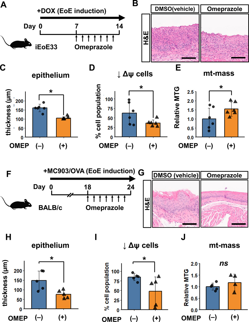
Background: Eosinophilic oesophagitis (EoE) is a food allergen-induced inflammatory disorder characterised by interleukin (IL)-13-mediated oesophageal inflammation and epithelial basal cell hyperplasia (BCH). The role of mitochondria in EoE pathogenesis remains elusive.

Design: Prompted by single cell transcriptomics data, we interrogated the role of mitochondria in EoE pathobiology using patient biopsies, EoE-mouse models and oesophageal epithelial cells grown in monolayer and three-dimensional (3D) organoid cultures treated with EoE-relevant cytokines. 3D organoids and EoE-bearing mice were treated with omeprazole-a proton-pump inhibitor used as first-line EoE therapy. We performed CRISPR (Clustered Regularly Interspaced Short Palindromic Repeats) interference in mouse organoids to identify the key mitochondrial regulatory genes whose depletion may lead to BCH. We analysed mitochondrial membrane potential, mass and superoxide production by flow cytometry, cellular oxygen consumption by respirometry, mitochondrial structures and perturbation of cellular energy homeostasis by immunoblotting. RESULTS : Mitochondrial dysfunction appeared to be a hallmark of EoE-related BCH where mitochondrial structural damage was associated with impaired oxidative respiratory capacity, elevation of mitochondrial superoxide and decreased adenosine triphosphate (ATP) production, as corroborated by activation of the adenosine monophosphate (AMP) -activated protein kinase and suppression of mammalian target-of-rapamycin signalling. Depletion of PGC1A, the master regulator of mitochondria biogenesis, recapitulated EoE-related BCH, suggesting that mitochondrial dysfunction drives BCH. Further, omeprazole alleviated mitochondrial damage and dysfunction in EoE-related BCH modelled in mice and patient-derived organoids. CONCLUSION: Mitochondrial dysfunction is tightly linked to perturbation of redox homeostasis in EoE-related BCH, which is promoted by IL-13 and reversible with omeprazole treatment.

Keywords: GASTROINTESTINAL IMMUNE RESPONSE; INFLAMMATORY DISEASES; OESOPHAGITIS.


Conflict of interest statement

Competing interests: None declared.

Figures

Figure 1
Figure 1. BCH lesions in EoE33 and doxycycline (DOX)-inducible iEoE33 mice contain epithelial cells with depolarized mitochondria.
EoE33 mice with chronic EoE via constitutive IL-13 expression and wild-type (WT) control (C57BL/6) mice were analyzed in (A-E). iEoE33 mice were fed with DOX-chow (experimental group) to induce acute EoE or normal chow (control) for 14 days and analyzed in (F-J). Upon euthanasia, H&E staining documented esophageal histology (A) and (F) and epithelial sheets were isolated for flow cytometry (B-E and G-J) of CD45-negative epithelial cells stained with MTDR and MTG to evaluate mitochondrial membrane potential (Δψ) and mitochondrial mass (mt-mass), respectively. Representative dot plots (B) and (G) are shown along with quantitation of cells with decreased mitochondrial membrane potential (↓ψΔ) in (C) and (H) and relative mitochondrial mass levels in (D) and (I). Percentage of cells with lower mt-mass in non-depolarized [ψΔ (+)] and depolarized [↓ψΔ] populations is shown in (E) and (J). *, p<0.05. ns; no significant difference. n=4 for both EoE33 and WT. n=6 for both DOX (−) and DOX (+). Scale bar, 100 μm.
Figure 2
Figure 2. BCH lesions display mitochondrial depolarization in murine EoE induced by SDS and MC903/OVA.
C57BL/6 mice received drinking water with 0.5% SDS (n=5, experimental group) or without (n=6, control) for 14 days in (A-D). BALB/c mice were exposed to MC903/OVA (n=5, experimental group) or vehicles only (n=5, control) in (E-H). Upon euthanasia, H&E staining documented esophageal histology (A) and (E) and epithelial sheets were isolated for flow cytometry (B-D and F-H). Representative dot plots (B) and (F) are shown along with quantitation of cells with decreased mitochondrial membrane potential (↓ψΔ) in (C) and (G) and relative mitochondrial mass levels in (D) and (H). *, p<0.05. ns; no significant difference. Scale bar, 100 μm.
Figure 3
Figure 3. BCH modeled in mouse esophageal 3D organoids demonstrates mitochondrial depolarization.
3D organoids grown from the C57BL/6 mouse esophagus were treated with or without 10 ng/mL IL-13 for up to 96h for H&E staining (A) and flow cytometry for mitochondrial depolarization (Δψ) and MTG (mt-mass) (B-E). Representative organoid morphology and flow cytometry dot plots at 96h (A) and (B) and quantification of cell populations with reduced Δψ at indicated time points (C). Percent of cells with lower mt-mass in non-depolarized and depolarized population at 96h is shown in (D). The relative MTG (mt-mass) levels at indicated time points in (E). The trapezium denotes the cell population with decreased Δψ. Scale bar, 100 μm. *, p<0.05.
Figure 4
Figure 4. IL-13 induces mitochondrial dysfunction in esophageal epithelial cells.
C57BL/6 mouse-derived esophageal 3D organoids (A) and (B) and primary esophageal epithelial monolayer culture (C-E) were treated with or without IL-13 at 10 ng/mL for 96h in (A) and (B) or 100 ng/mL for 48h in (C-E) and subjected to mitochondria-related assays. In (A), cells were incubated with MitoSOX for flow cytometry to measure mitochondrial superoxide level. In (B), whole cell lysates were used to measure the ATP level per 1 × 10⁶ cells. In (C-E), respirometry was done under baseline and stressed conditions with representative oxidative respiration (baseline and stressed OCR) and glycolysis (stressed ECAR) shown in (D) and (E). Oligomycin (O), FCCP (F), and rotenone along with antimycin A (RA) were used at indicated time points to disrupt mitochondrial function and electron flow through the electron transport chain, inducing stress conditions. *, p<0.05; ns, not significant vs. IL-13 (−).
Figure 5
Figure 5. IL-13 activates AMPK and suppresses mTORC1 signaling in esophageal 3D organoids.
C57BL/6 mouse-derived esophageal 3D organoids were treated with or without IL-13 for 96h and subjected to immunoblotting. Representative blots for indicated proteins with densitometry (A-F). Actin served as a loading control. Mitochondrial depolarization in 3D organoids treated with rapamycin (RM, 100 nM) or DMSO (vehicle control) in the presence or absence of IL-13 for 96 hours (G). Mitochondrial depolarization in 3D organoids receiving AS1517499 (AS, 400 nM) or vehicle control (DMSO), administered 24 hours before IL-13 exposure and maintained during the subsequent 16-hour IL-13 treatment (H). *, p<0.05, ns; no significant difference.
Figure 6
Figure 6. IL-13 disrupts mitochondrial network architecture in esophageal epithelial cells.
C57BL/6 mouse-derived esophageal primary esophageal epithelial monolayer culture was treated with 10 ng/mL IL-13 for 48h and stained with MitoTracker Red CMXRos dye to visualize mitochondria by confocal microscopy. Representative images are shown with areas enlarged to highlight a filamentous mitochondrial network in untreated cells and the fragmented network with ring-shaped structures (a hallmark of damaged mitochondria, arrows) in IL-13-treated cells in (A). The captured images were subjected to mitochondrial network analysis to quantitate branches and junctions as demonstrated in (B-D). *, p<0.05; n=4. Scale bar, 10 μm.
Figure 7
Figure 7. Pgc1a depletion in esophageal epithelial cells causes BCH-like changes with mitochondrial damage.
Following CRISPRi-mediated Pgc1a knockdown, mouse esophageal epithelial cells were subjected to organoid formation for morphological assessment of BCH-like changes by H&E staining, SOX2 staining, and mRNA expression of Sox2 by qRT-PCR in (A-D) and flow cytometry for MTDR (Δψ) in (E), or grown in monolayer culture for quantitative evaluation of the mitochondrial network in (F-H). Cells with sgRNA targeting Pgc1a (sgPgc1a) were compared to those with non-targeting control sgRNA (sgNT). Scale bar, 100 μm in (A) and (B), 10 μm. in (F). *, p<0.05.
Figure 8
Figure 8. Omeprazole treatment alleviates BCH in EoE-bearing mice.
iEoE33 mice exhibiting DOX-induced EoE were given daily either omeprazole (OMEP) (n=6) or DMSO (vehicle control) (n = 6) via intragastric gavage and sacrificed (A) to evaluate histopathology by H&E staining (B) and (C) and flow cytometry for MTDR (Δψ) (D) and mt-mass (E) in the esophageal epithelium. Epithelial thickness was measured to assess BCH in (C). MC903/OVA treated mice were given daily either omeprazole (OMEP) (n=5) or DMSO (vehicle control) (n = 5) via intragastric gavage and sacrificed (F) to evaluate histopathology by H&E staining (G and H) and flow cytometry for MTDR (Δψ) (I) and mt-mass (J) in the esophageal epithelium. Epithelial thickness was measured to assess BCH in (C) and (H). Scale bar, 100 μm in (B) and (G). *, p<0.05 in (C-E).
Figure 9
Figure 9. Omeprazole prevents IL-13 from inducing BCH-like changes in mouse esophageal 3D organoids.
C57BL/6 mouse-derived esophageal 3D organoids were treated with or without 10 ng/mL IL-13 concurrent with or without with 200 μM omeprazole (OMEP) or DMSO (vehicle control) for four days to evaluate morphological changes by H&E staining (A) and p-STAT6 immunofluorescence (B). p-STAT6+ cells were quantitated in (C). Scale bar, 100 μm. *, p<0.05.
Figure 10
Figure 10. Omeprazole restores mitochondrial damage and mTORC1 signaling in mouse esophageal 3D organoids.
C57BL/6 mouse-derived esophageal 3D organoids were treated with or without 10 ng/mL IL-13 concurrent with or without with 200 μM omeprazole (OMEP) or DMSO (vehicle control) for four days and subjected to flow cytometry for MTDR (Δψ) (A), mt-mass (B), and mitochondrial superoxide (C), and immunoblot analysis of whole cell lysates (D-F). Representative blots are shown for indicated proteins (D) with densitometry in (E) and (F). Actin served as a loading control. Cells transfected with sgRNA targeting Pgc1a (sgPgc1a) or non-targeting control sgRNA (sgNT) were treated with or without 10 ng/mL IL-13 for four days and subsequently analyzed by MTDR (Δψ) (G). Pgc1a mRNA expression by qRT-PCR in (H). *, p<0.05, ns; no significant difference.
Figure 11
Figure 11. Patient biopsies display mitochondrial depolarization and correlate with EoE inflammation and the tissue IL-13 level.
Endoscopic esophageal biopsies were dissociated into single cells and subjected to flow cytometry for MTDR (Δψ) (A) and (B) and MTG (mt-mass) (C) and (D) within the non-immune (CD45-) cell population. Pediatric subjects consisted of those with normal esophageal mucosa (n=5), inactive EoE (n=5), and active EoE (n=5) in (A) and (B). Adults consisted of those with normal esophageal mucosa (n=5), inactive EoE (n=5) and active EoE (n=6) in (C) and (D). *, p<0.05; ns, not significant. The percentage of cells with decreased Δψ (↓Δψ cells) in biopsy samples (A-D) was correlated with tissue eosinophil counts [number of cells per high power field (HPF)] and the relative IL-13 mRNA expression level determined by qRT-PCR (GAPDH internal control) in (E-G).
Figure 12
Figure 12. Omeprazole restores mitochondrial dysfunction in patient-derived organoids.
Patient-derived esophageal 3D organoids were treated with or without 10 ng/mL IL-13 concurrent with or without with 200 μM omeprazole (OMEP) or DMSO (vehicle control) for four days and subjected to flow cytometry for MTDR (Δψ) (A) and mitochondrial superoxide (B). *, p<0.05; ns, not significant.
Figure 13
Figure 13. Graphical abstract of the proposed model.
The pathogenesis of EoE involves epithelial barrier defect, exposure to food allergens, and IL-13-mediated inflammation, culminating in BCH linked to mitochondrial dysfunction. PGC1A, the master regulator of mitochondrial biogenesis, serves to maintain the esophageal epithelial redox homeostasis to prevent BCH. PPIs like omeprazole (OMEP) prevent mitochondrial damage and BCH, possibly through mechanisms like downregulation of IL-13Rα1, a subunit of the IL-13 receptor, and upregulation of peroxisome proliferator-activated receptor gamma (PPARγ), a key binding partner of PGC1A, that regulates cell metabolism.

References

    1. Doyle AD, Masuda MY, Pyon GC, et al. Detergent exposure induces epithelial barrier dysfunction and eosinophilic inflammation in the esophagus. Allergy. 2023;78:192–201. doi: 10.1111/all.15457 - DOI - PMC - PubMed
    1. Wright BL, Masuda MY, Ortiz DR, et al. Allergies Come Clean: The Role of Detergents in Epithelial Barrier Dysfunction. Curr Allergy Asthma Rep. 2023;23:443–51. doi: 10.1007/s11882-023-01094-x - DOI - PMC - PubMed
    1. Marella S, Sharma A, Ganesan V, et al. IL-13-induced STAT3-dependent signaling networks regulate esophageal epithelial proliferation in eosinophilic esophagitis. J Allergy Clin Immunol. 2023;152:1550–68. doi: 10.1016/j.jaci.2023.07.021 - DOI - PMC - PubMed
    1. Marone G, Granata F, Pucino V, et al. The Intriguing Role of Interleukin 13 in the PathophysiologyofAsthma. FrontPharmacol.2019;10:1387.doi: 10.3389/fphar.2019.01387 - DOI - PMC - PubMed
    1. Rochman M, Xie YM, Mack L, et al. Broad transcriptional response of the human esophageal epithelium to proton pump inhibitors. J Allergy Clin Immunol. 2021;147:1924–35. doi: 10.1016/j.jaci.2020.09.039 - DOI - PMC - PubMed

LinkOut - more resources