. 2025 Jul 23;16(1):551.
doi: 10.1038/s41419-025-07794-8.

UBE4B promotes gastric cancer proliferation and metastasis by mediating FAT4 ubiquitination and degradation

Affiliations

UBE4B promotes gastric cancer proliferation and metastasis by mediating FAT4 ubiquitination and degradation

Kaini Wu et al. Cell Death Dis. .

Abstract

The ubiquitin‒proteasome system (UPS), an intracellular protein degradation pathway, plays an important role in regulating tumorigenesis and development. Ubiquitination factor E4B (UBE4B/UFD2) has been shown to be associated with the development of several cancers. The aim of this study was to reveal the functional significance of UBE4B in gastric cancer (GC) development and its important mechanism. Bioinformatics analysis, immunohistochemistry (IHC), western blotting, and real-time PCR were performed to detect UBE4B expression in human GC samples and GC cell lines and a mouse xenograft tumour model was established. Our investigation revealed that UBE4B is highly expressed in GC and promotes the proliferation, migration and invasion of GC cells. The quantitative Tandem Mass Tag (TMT) analysis revealed that FAT oncogenic homologue 4 (FAT4) is a downstream gene of UBE4B. Western blot experiments and transmission electron microscopy (TEM) results for biological samples revealed that UBE4B inhibits autophagy in GC cells and directly binds to and degrades FAT4 through ubiquitination. These results suggest that UBE4B can inhibit autophagy and promote GC progression by mediating FAT4 ubiquitination and degradation, and our findings provide a new potential therapeutic target for GC management.


Conflict of interest statement

Competing interests: The authors declare no competing interests. Ethical approval: Inclusion of human participants, and use of human data and human tissue in this study were approved by the Ethics Committee of the First Affiliated Hospital of Nanchang University. The use of animals in this study was approved by the animal research committee in the First Affiliated Hospital of Nanchang University.

Figures
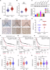
Fig. 1
Fig. 1. UBE4B was elevated in gastric cancer.
A The real-time PCR assay of the mRNA expression of UBE4B in gastric cancer (GC) tissues and normal tissues from the TCGA database. B The real-time PCR assay of the mRNA expression of UBE4B in GC tissues and normal tissues from the GEO database. C The basic UBE4B protein level in different GC cell types. D Immunohistochemistry (IHC) staining for UBE4B in GC and adjacent normal tissue. Bar length: 50 μm. E IHC score analyzed the expression levels of UBE4B in GC and adjacent normal tissue. FH The Kaplan–Meier plot of Overall survival (OS), progression-free survival (FP), and post-progression survival (PPS) by the expression of UBE4B in the GC patients, the data was carried out from the TCGA database. IK Clinicopathologic features of UBE4B expression and N-stage, M-stage, and stage in GC patients, the data was carried out from the GEO database.
Fig. 2
Fig. 2. UBE4B promoted the proliferation, migration, and invasion of GC cell lines.
A Western blot analysis of UBE4B protein levels and Real-time PCR analysis of UBE4B mRNA levels in knockdown cell line (MKN45) and overexpression cell line (AGS). B Colony formation assay was used to detect the proliferation of knockdown UBE4B and control GC cells, and overexpressing UBE4B and control GC cells. C Invasion and migration assay were performed to detect the proliferation of knockdown UBE4B and control MKN45 cells. Bar length: 100 μm. D Invasion and migration assays were performed to detect the proliferation of overexpression UBE4B and control AGS cells. Bar length: 100 μm. E The CCK-8 assay was performed to detect the proliferation of knockdown UBE4B and control MKN45 cells. F The CCK-8 assay was performed to detect the proliferation of overexpression UBE4B and control AGS cells.
Fig. 3
Fig. 3. UBE4B mediates the degradation of FAT4.
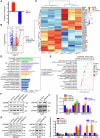
A Mass spectroscopic analysis of Up-regulated and down-regulated protein. B Volcano plot showing differentially expressed proteins (P < 0.05, FC < 1/1.2 & FC > 1.2). C Heatmap shows a differentially expressed gene. D KEGG enrichment analysis top 20. E KEGG Level 3 shows differential protein distribution. F Western blot detected FAT4 protein in MKN45 or AGS cells after transfecting cells with three independent siRNA or UBE4B overexpression plasmid. G Western blot detected P62 and LC3II/I protein in MKN45 or AGS cells after transfecting cells with three independent siRNA or UBE4B overexpression plasmid.
Fig. 4
Fig. 4. UBE4B mediates the degradation of FAT4 through ubiquitination.
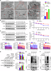
A Transmission electron microscopy observation of autophagosomes in UBE4B-knockdown MKN45 cells. B, C MKN45 cells transfected with two independent siUBE4B were treated with 20 μM cycloheximide (CHX) for 0, 2, 4, 6, 8, and 12 h, and the UBE4B and FAT4 protein levels were detected by Western blot. D Western blotting was conducted to measure FAT4 protein level after treatment with 20 μM MG132 in knockdown UBE4B and control GC cells. E, F Western blotting was conducted to measure the level of ubiquitin when knockdown or overexpression UBE4B.
Fig. 5
Fig. 5. UBE4B interacts with FAT4 through ubiquitination.
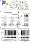
A Schematic representation of the docking structure of protein molecules. Blue color indicates UBE4B protein and yellow color indicates FAT4 protein. The key interacting residues are highlighted in red font. B Segment plasmid of UBE4B. C, D Endogenous UBE4B proteins were immunoprecipitated with an anti-UBE4B antibody and then analyzed by immunoblotting. Endogenous FAT4 proteins were immunoprecipitated with anti-FAT4 antibodies and then analyzed by immunoblotting. The IgG antibody was used as the control. E HEK-293T cells infected endogenous overexpression of FAT4 lentivirus and HA-UBE4B plasmid were subject to immunoprecipitation with anti-UBE4B and anti-FAT4 antibodies. F HEK-293T cells infected endogenous overexpression of FAT4 lentivirus were transfected with HA-UBE4B or several UBE4B mutants. A coimmunoprecipitation assay was performed to detect the interaction between UBE4B and FAT4 protein. G HEK-293T cells infected with endogenous overexpression of FAT4 lentivirus were transfected with HA-UBE4B or wild-type Ub, Ub-K6R, K11R, K27R, K29R, K33R, K48R, and K63R plasmid. Cell lysates were assayed for ubiquitination, and the ubiquitination level of FAT4 was detected. H HEK-293T cells infected with endogenous overexpression of FAT4 lentivirus were transfected with HA-UBE4B or wild-type Ub, Ub-K6R, K33R and K6R/K33R plasmid. Cell lysates were assayed for ubiquitination, and the ubiquitination level of FAT4 was detected.
Fig. 6
Fig. 6. The carcinogenic effect of UBE4B is dependent on the degradation of FAT4.
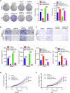
A Colony formation assay was performed to detect the proliferation of GC cells. B Invasion and migration assays were performed to detect the proliferation of MKN45 cells. Bar length: 100 μm. C Invasion and migration assays were performed to detect the proliferation of AGS cells. Bar length: 100 μm. D CCK-8 assay was used to detect the proliferation of MKN45 cells. E CCK-8 assay was used to detect the proliferation of AGS cells.
Fig. 7
Fig. 7. UBE4B downregulates FAT4 to promote GC cell proliferation in vivo.
A MKN45 cells with/without shUBE4B were injected in nude mice. Representative images of xenograft tumors from mice bearing MKN45-shNC and MKN45-shUBE4B cells. B Tumor weights were calculated for each mouse in both groups (n = 6, Student’s t test). C Tumor volume was measured every 5 days (n = 6, Student’s t test). D IHC scores were analyzed for the expression levels of UBE4B, FAT4, and Ki67 in both groups (NC vs UBE4B-OE, n = 15, Student’s t test). E, F UBE4B, FAT4, and Ki67 expression in the tumor of nude mice were detected by IHC. Bar length: 50 μm. G MKN45 cells with/without shUBE4B or with/without FAT4 were injected in nude mice. Representative images of xenograft tumors from mice bearing MKN45-shNC+shNC, MKN45-shUBE4B+shNC, and MKN45-shUBE4B+shFAT4 cells. H Tumor weights were calculated for each mouse in the three groups (n = 10, One-way ANOVA). I Tumor volume was measured every 3 days (n = 10, One-way ANOVA). J IHC scores were analyzed for the expression levels of UBE4B, FAT4, and Ki67 in the three groups (NC + NC vs KD-UBE4B + NC vs KD-UBE4B + KD-FAT4, n = 15, One-way ANOVA).
Fig. 8
Fig. 8. UBE4B was highly expressed in clinical GC samples, and patients had a poor prognosis, in contrast to FAT4.
A Representative image of IHC staining of tumor microarrays from GC patients. Bar length: 50 μm. B IHC scores of UBE4B level between GC tissue and adjacent normal tissue. C IHC scores of FAT4 level between GC tissue and adjacent normal tissue. D The Kaplan-Meier plot of overall survival by the expression of UBE4B in tumor microarrays related information (n = 66 (high-expression) vs 28 (low-expression)). E The Kaplan-Meier plot of overall survival by the expression of FAT4 in tumor microarrays related information (n = 46 (high-expression) vs 48 (low-expression)).
Fig. 9
Fig. 9. Scheme for the regulatory mechanism of UBE4B on FAT4.
Created with BioRender.com.

References

    1. Bray F, Laversanne M, Sung H, Ferlay J, Siegel RL, Soerjomataram I, et al. Global cancer statistics 2022: GLOBOCAN estimates of incidence and mortality worldwide for 36 cancers in 185 countries. CA Cancer J Clin. 2024;74:229–63. - PubMed
    1. Herrero R, Parsonnet J, Greenberg ER. Prevention of gastric cancer. Jama. 2014;312:1197–8. - PubMed
    1. Chen L, Liu S, Tao Y. Regulating tumor suppressor genes: post-translational modifications. Signal Transduct Target Ther. 2020;5:90. - PMC - PubMed
    1. Wang Y, Le WD. Autophagy and ubiquitin-proteasome system. Adv Exp Med Biol. 2019;1206:527–50. - PubMed
    1. Aliabadi F, Sohrabi B, Mostafavi E, Pazoki-Toroudi H, Webster TJ. Ubiquitin-proteasome system and the role of its inhibitors in cancer therapy. Open Biol. 2021;11:200390. - PMC - PubMed

Substances