. 2025 Jul;57(7):1579-1592.
doi: 10.1038/s12276-025-01503-3. Epub 2025 Jul 23.

piENOX2 regulates ALKBH5-mediated Itga4 m6A modification to accelerate the progression of rheumatoid arthritis

Affiliations

piENOX2 regulates ALKBH5-mediated Itga4 m6A modification to accelerate the progression of rheumatoid arthritis

Naibo Feng et al. Exp Mol Med. 2025 Jul.

Abstract

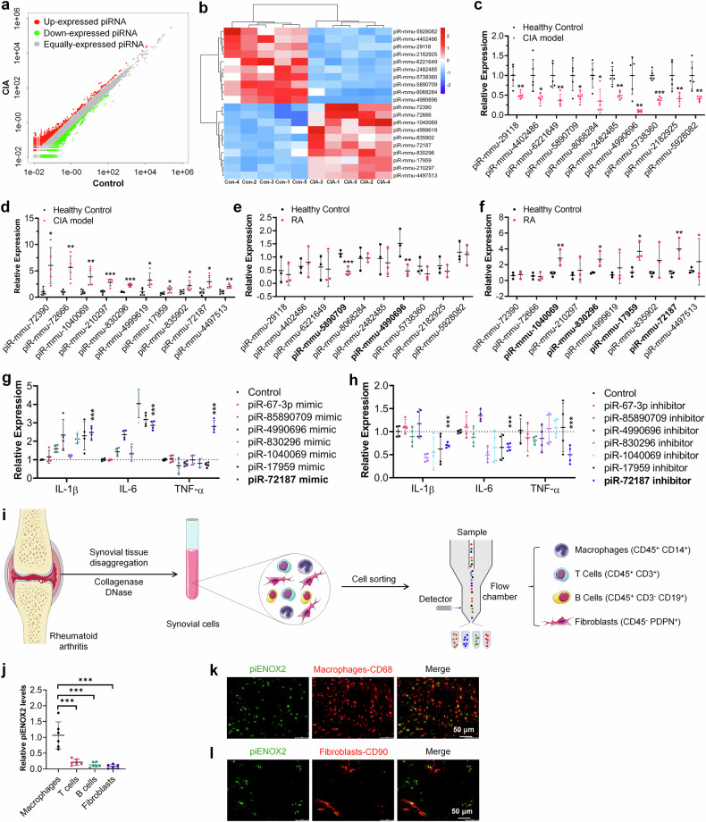
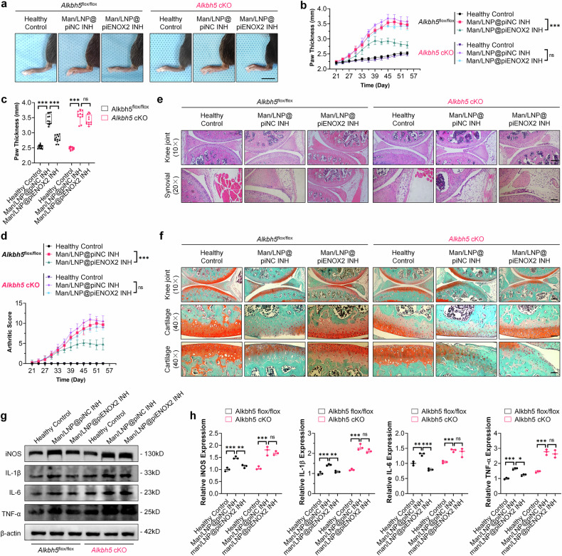
Rheumatoid arthritis (RA) is a chronic autoimmune disorder characterized by synovitis and presenting as symmetrical arthritis that primarily affects the small joints of the limbs. PIWI-interacting RNAs, a class of small noncoding RNAs, have garnered significant attention due to their critical involvement in various pathological conditions, including reproductive diseases, cancers and other disorders. Here we observe elevated levels of macrophage-derived piENOX2 in the synovial tissues of both patients with RA and mice with collagen-induced arthritis (CIA). It was found that transfection with a piENOX2 mimic promoted M1 macrophage polarization, while a piENOX2 inhibitor facilitated M2 polarization. In vivo, a piENOX2 inhibitor significantly alleviated disease progression, reduced systemic inflammation and preserved the integrity of articular cartilage in CIA mice. Mechanistic analyses indicated that the piENOX2 effects were due to its targeting Alkbh5 mRNA for degradation. In a Alkbh5 conditional-knockout mouse model of CIA, the therapeutic effects of a piENOX2 inhibitor, including inflammation suppression and cartilage protection, were reduced compared with control mice. A comprehensive analyses using methylated RNA immunoprecipitation sequencing and methylated RNA immunoprecipitation and quantitative PCR revealed that piENOX2 regulated ALKBH5-mediated m6A modification of Itga4 mRNA, thereby influencing macrophage polarization through the PI3K-AKT signaling pathway. These findings provide important insights into the complex roles of PIWI-interacting RNAs in RA progression and indicate potential avenues for therapeutic intervention.


Conflict of interest statement

Competing interests: The authors declare no competing interests.

Figures

Fig. 1
Fig. 1. Macrophage-derived piENOX2 plays an important role in the progression of RA.
a The piRNA expression in each sample was analyzed using the Illumina HiSeq 2500 system (N = 5). The CIA mouse model was constructed, and hind-limb knee joint synovial tissue samples were collected at predetermined time points for total RNA extraction. bd A cluster analysis and expression profiling of the top ten human–mice homologous piRNAs with the most significant upregulation and downregulation. e, f RT–qPCR was used to detect the expression of these piRNAs in the synovial tissue of patients with RA; in the LPS-induced inflammation model, piRNA mimics (g) and inhibitors (h) were used to overexpress and inhibit the expression of corresponding piRNAs, respectively, and RT–qPCR was performed to measure the levels of TNF, IL-1β and IL-6. The synovial tissue samples from the affected joints of CIA mice were isolated, and single-cell suspensions were prepared through enzymatic hydrolysis. i, The key cell populations in the synovial tissue, including synovial macrophages (CD45+CD14+), T cells (CD45+CD3+), B cells (CD45+CD3CD19+) and fibroblasts (CD45PDPN+), were sorted and collected via flow cytometry. j, RT–qPCR analysis of piENOX2 expression in individual cell populations (N = 6). k The distribution and colocalization of piENOX2 and synovial macrophages (CD68+) in the synovial tissue of patients with RA were observed using FISH. l The distribution and colocalization of piENOX2 and fibroblasts (CD90+) in the synovial tissue of patients with RA were observed using FISH. * P< 0.05, ** P< 0.01, *** P< 0.001.
Fig. 2
Fig. 2. piENOX2 is upregulated in the synovial tissue of patients with RA and is involved in the regulation of macrophage polarization.
a FISH observation of piENOX2 expression and distribution in the synovial tissue of patients with RA and control patients. b Semiquantitative analysis of piENOX2 fluorescence intensity in different samples using ImageJ (arbitrary units (AU)). c RT–qPCR detection of piENOX2 expression in the synovial tissue of patients with RA and controls (Ncontrol = 20, NRA = 23). d A Pearson correlation analysis was performed to evaluate the association between DAS-28 scores and piENOX2 expression levels (N = 24). e, f Flow cytometry (e) and quantitative analysis (f) of the regulatory effects of piENOX2 MIM and piENOX2 INH on IL-4-induced M2 polarization (N = 3). g, h Western blotting (g) showing the regulatory effects of piENOX2 MIM and piENOX2 INH on the expression of M2 macrophage markers and their secreted cytokines, including ARG1, TGF-β and IL-10, with semiquantitative analysis (h) performed using ImageJ (N = 3). i, j Flow cytometry (i) and quantitative analysis (j) of the regulatory effects of piENOX2 MIM and piENOX2 INH on LPS-induced M1 polarization (N = 4). k, l Western blot analysis (k) of the regulatory effects of piENOX2 MIM and piENOX2 INH on the expression of M1 macrophage markers and their secreted cytokines, including iNOS, IL-1β, IL-6 and TNF, with semiquantitative analysis (l) performed using ImageJ (N = 3). *P < 0.05, **P < 0.01, ***P < 0.001.
Fig. 3
Fig. 3. Man/LNP@piENOX2 INH effectively alleviates disease progression in CIA mice.
a The construction of Man/LNP@piENOX2 INH. b TEM image of Man/LNP@piENOX2 INH. c The tissue distribution of ICG-labeled Man/LNP@piENOX2 INH in CIA mice, observed via small animal in vivo imaging at predetermined time points following tail vein injection. d Photographs of hind paws in each group of mice; scale bar, 10 mm. e Trend of changes in hind-paw thickness over the treatment period. f Final measured hind-paw thickness in each group. g Trends in arthritis scores of mice in each group throughout the treatment period. h A hematoxylin and eosin staining showing joint morphology and synovial infiltration in each group of mice; scale bars, 200 μm at 10× and 100 μm at 20× magnification. i Safranin-O and Fast Green staining indicating joint cartilage damage in each group of mice; scale bars, 200 μm at 10× and 50 μm at 40× magnification. j, k The protein expression levels of iNOS, IL-1β, IL-6 and TNF in the synovial tissue of each group of mice, detected by western blotting (j), with semiquantification (k) using ImageJ (N = 3). *P < 0.05, **P < 0.01, ***P < 0.001.
Fig. 4
Fig. 4. piENOX2 regulates macrophage polarization by targeting the degradation of Alkbh5 mRNA.
a Base complementarity between piENOX2 (human piRNA: piR-has-13893) and Alkbh5 mRNA in humans and mice. b A simulation of the binding between piENOX2, PIWI and Alkbh5 mRNA using visual molecular docking. c, d RT–qPCR (c) and western blots (d) showing the effects of piENOX2 MIM and piENOX2 INH on Alkbh5 mRNA and protein expression (N = 3). e, f Flow cytometry (e) and quantitative analysis (f) of the effect of Alkbh5 knockdown on piENOX2 INH-mediated promotion of M2 macrophage polarization (N = 3). g, h Flow cytometry (g) and quantitative analysis (h) of the effect of Alkbh5 overexpression on piENOX2 MIM-mediated promotion of M1 macrophage polarization (N = 4). i, j The evaluation of the effect of Alkbh5 overexpression or knockdown on piENOX2-regulated M2 macrophage polarization by western blot (i), with semiquantification (j) using ImageJ (N = 3). k, l The evaluation of the effect of Alkbh5 overexpression or knockdown on piENOX2-regulated M1 macrophage polarization by western blotting (k), with semiquantification (l) using ImageJ (N = 3). *P < 0.05, **P< 0.01, ***P< 0.001.
Fig. 5
Fig. 5. Knockout of Alkbh5 blocks the effect of Man/LNP@piENOX2 INH on delaying disease progression in CIA mice.
a An observation of swelling and redness in the hind paws of CIA mice in each treatment group at predetermined time points following treatment with different drugs; scale bar, 10 mm. b, c The trend of hind-paw thickness changes over the entire treatment period (b) and final measurement of hind-paw thickness in each group (c) (N = 6). d The trends in arthritis scores of mice in each group throughout the treatment period (N = 6). e Hematoxylin and eosin staining showing joint morphology and synovial infiltration in each group of mice; scale bars, 200 μm at 10× and 100 μm at 20× magnification. f Safranin-O and Fast Green staining indicating joint cartilage damage in each group of mice; scale bars 200 μm at 10× and 50 μm at 40× magnification. g, h Protein expression levels of iNOS, IL-1β, IL-6 and TNF in the synovial tissue of each group of mice, detected by western blotting (g), with semiquantification (h) using ImageJ (N = 3). A statistical analysis was conducted on data from day 54. ns, no significance; *P < 0.05, **P < 0.01, ***P < 0.001.
Fig. 6
Fig. 6. piENOX2 regulates ALKBH5-mediated m6A modification of Itga4 mRNA, thereby influencing macrophage polarization through the PI3K–AKT signaling pathway.
a An Integrated Genome Viewer visualization of the methylation level of Itga4 mRNA. b meRIP–qPCR detection of Itga4 m6A levels following treatment with piENOX2 MIM and piENOX2 INH (N = 3). c RT–qPCR detection of Itga4 mRNA expression after treatment with piENOX2 MIM and piENOX2 INH (N = 3). d Western blotting of ITGA4 protein expression after treatment with piENOX2 MIM and piENOX2 INH. e, f Western blotting (e) showing the effects of the effects of Itga4 knockdown on activation of the PI3K–AKT signaling pathway by piENOX2 INH, with semiquantification (f) using ImageJ (N = 3). g Flow cytometry analysis of the effects of Itga4 knockdown or blocking the PI3K–AKT signaling pathway on piENOX2 INH-promoted M2 macrophage polarization (N = 4). h, i Western blotting (h) showing the effects of Itga4 knockdown or blocking the PI3K–AKT signaling pathway on piENOX2 INH-promoted M2 macrophage polarization, with semiquantification (i) using ImageJ (N = 3). j Flow cytometry analysis of the effects of Itga4 knockdown or blocking the PI3K–AKT signaling pathway on piENOX2 INH-inhibited M1 macrophage polarization (N = 4). k, l Western blotting (k) showing the effects of Itga4 knockdown or blocking the PI3K–AKT signaling pathway on piENOX2 INH-inhibited M1 macrophage polarization, with semiquantification (l) using ImageJ (N = 3). ns no significance; **P< 0.01, ***P < 0.001.
Fig. 7
Fig. 7. Schematic diagram of the mechanism by which piENOX2 accelerates RA disease progression.
Under physiological conditions, ALKBH5 stabilizes Itga4 mRNA expression through demethylation, subsequently activating the PI3K–AKT signaling pathway. This regulatory mechanism promotes macrophage M2 polarization, maintaining immune homeostasis. In RA, piENOX2 production is significantly elevated. piENOX2 selectively targets Alkbh5 mRNA for degradation, thereby inhibiting ALKBH5 protein expression. This disruption leads to increased m6A modification on Itga4, reducing its mRNA stability and decreasing ITGA4 expression. As a result, ITGA4-mediated activation of the PI3K–AKT pathway is inhibited, promoting macrophage M1 polarization. This cascade of events ultimately contributes to the onset and progression of RA.

Similar articles

References

    1. Smith, M. H. & Berman, J. R. What is rheumatoid arthritis? Jama327, 1194 (2022). - PubMed
    1. Smolen, J. S. et al. EULAR recommendations for the management of rheumatoid arthritis with synthetic and biological disease-modifying antirheumatic drugs: 2022 update. Ann. Rheum. Dis.82, 3–18 (2022). - PubMed
    1. Aravin, A. et al. A novel class of small RNAs bind to MILI protein in mouse testes. Nature442, 203–207 (2006). - PubMed
    1. Girard, A., Sachidanandam, R., Hannon, G. J. & Carmell, M. A. A germline-specific class of small RNAs binds mammalian Piwi proteins. Nature442, 199–202 (2006). - PubMed
    1. Grivna, S. T., Beyret, E., Wang, Z. & Lin, H. A novel class of small RNAs in mouse spermatogenic cells. Genes Dev.20, 1709–1714 (2006). - PMC - PubMed

Substances

LinkOut - more resources