. 2025 Jul 23;15(1):26767.
doi: 10.1038/s41598-025-12331-w.

Topical ophthalmic administration of the antiangiogenic peptide VIAN-c4551 protects against experimental diabetic macular edema

Affiliations

Topical ophthalmic administration of the antiangiogenic peptide VIAN-c4551 protects against experimental diabetic macular edema

Elva Adán-Castro et al. Sci Rep. .

Abstract

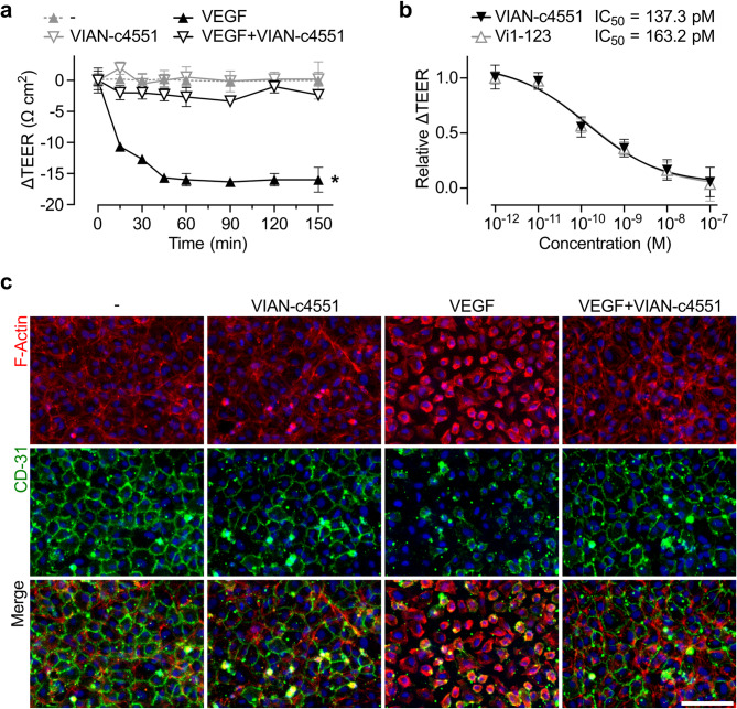
Increased angiogenesis and vascular permeability are hallmarks of microvascular retinal diseases such as diabetic retinopathy and diabetic macular edema (DME). Periodic intravitreal injections of inhibitors of the vascular endothelial growth factor (VEGF) are first-line therapy, but their invasiveness and associated risks often lead to poor compliance and outcomes. Here, we investigate VIAN-c4551, a highly potent antiangiogenic cyclic heptapeptide, as a non-invasive topical ophthalmic alternative to the current standard of care for DME. VIAN-c4551 demonstrated high potency (IC50 = 137 pM) to inhibit the permeability of human umbilical vein endothelial cell monolayers induced by VEGF. VIAN-c4551 eye drops potently (0.005% minimum effective dose) prevented, for up to 24 h, the retinal vascular leakage induced by VEGF injected intravitreally and reversed the increased retinal vascular permeability due to diabetes in rats and mice. VIAN-c4551 exhibited high penetrability across MDCK epithelium and, after a single eye drop in rabbits, reached the vitreous and the retina-choroid at concentrations several orders of magnitude above its IC50 (Cmax ~239 nM and ~ 6.7 µM, respectively, after 6 h) that lasted at least 24 h. In conclusion, VIAN-c4551 is a non-invasive, once-a-day potential intervention for preventing and reversing retinal vascular leakage in DME and other vascular retinopathies and preserving sight.

Keywords: Anti-angiogenesis; Diabetic macular edema; Diabetic retinopathy; Efficacy; Eye drops; Pharmacokinetics; Therapeutic peptide; Vascular permeability; Vasoinhibin analog.


Conflict of interest statement

Declarations. Competing interests: JPR, MZ, TB, JT, GME, and CC are inventors of a submitted patent application (WO/2021/098996), which is owned by the Universidad Nacional Autónoma de México (UNAM) and JT. JPR is the CEO and founder of VIAN Therapeutics Inc. MZ and CC are consultants for VIAN Therapeutics. Inc.

Figures

Fig. 1
Fig. 1
VIAN-c4551 inhibits VEGF-induced permeability of HUVEC monolayers. (a) Changes in the trans-endothelial electrical resistance (ΔTEER) of HUVEC monolayers treated or not with 100 nM VIAN-c4551 throughout a 150-minute incubation period without (-) or with VEGF (50 ng mL− 1). Values are means ± SD, n = 3, *P < 0.006 vs. (-) for all time points except 0 (2-way ANOVA repeated measures, Dunnett’s). (b) Dose-response inhibition of the VEGF-induced reduction of ΔTEER at 90 min by different concentrations of VIAN-c4551 or Vi1-123. The IC50 of VIAN-c4551 and Vi1-123 are indicated. ΔTEER is relative to that of VEGF alone. Values are means ± SD, n = 9, and the curve represents a nonlinear regression analysis of four parameters (54 points, r2 ≥ 0.92). (c) Representative fluorescence of HUVEC monolayers stained for F-actin and CD31 pretreated with or without VIAN-c4551 for 1 h followed by VEGF (200 ng mL− 1) for 30 min. Nuclei were counterstained with Hoechst 33,342. Scale bar = 100 μm.
Fig. 2
Fig. 2
VIAN-c4551 transit across MDCK epithelial cells. (a) Percentage of transit across an MDCK monolayer of two concentrations (0.4 and 4 nM) of VIAN-c4551 or Vi1-123 after 2 h relative to their transit of both compounds in the absence of MDCK monolayers. Bars indicate mean ± SD, n ≥ 6. Individual values are shown. (b) Permeability coefficient (Papp) of VIAN-c4551 and Vi1-123.
Fig. 3
Fig. 3
VIAN-c4551 eye drops potently inhibit VEGF-induced retinal vascular permeability for up to 24 h. Retinal vascular permeability in rats (a) and mice (b) treated with a single eye drop containing 0.5% VIAN-c4551 or vehicle administered one hour before the intravitreal injection of 250 ng of VEGF or PBS. (c) Dose-response inhibition of eye drops containing different concentrations of VIAN-c4551. (d) Duration of the effect of a single 0.5% VIAN-c4551 eye drop administered at different times before the intravitreal injection of VEGF. Quantification of Evans blue-stained albumin extravasation was performed two hours after VEGF administration. Values are means ± SD. Individual eye values are shown. Sample size (n) was 15–22 (a), 7–16 (b), 9–23 (c), and 5–27 (d). #P < 0.002 vs. no VEGF (-), *P < 0.033, **P < 0.002, ***P < 0.001 vs. Veh (-), 2-way ANOVA, Šidák’s (a, b), 1-way ANOVA, Dunnett’s (c, d).
Fig. 4
Fig. 4
Visualization of VIAN-c4551 eye drop inhibition of VEGF-induced retinal vascular leakage. Representative fluorescein angiography images of flat-mounted retinas from rats treated with Veh or VIAN-c4551 eyedrops 4 h before intravitreal injection of PBS or VEGF and euthanized 2 h post-intravitreal injections. Retinal vasculature was visualized by FITC-dextran perfusion 1 h after intravitreal injection of VEGF and 1 h of circulation. Vascular leakage areas (arrowheads) and optic nerve (ON) are indicated. Scale bar = 1 mm.
Fig. 5
Fig. 5
VIAN-c4551 inhibits diabetes-induced retinal vascular permeability. Retinal vascular permeability in nondiabetic (NDB) and 6-week diabetic (DB) rats (a) and mice (b) treated daily for 5 days with a single eye drop containing 0.5% VIAN-c4551 or vehicle. The extravasation of Evans blue-stained albumin evaluated retinal vascular permeability 24 h after the last eye drop. Values are means ± SD. Individual eye values are shown. #P < 0.002 vs. NDB, **P < 0.002, ***P < 0.001 vs. Veh, (2-way ANOVA, Šidák’s).
Fig. 6
Fig. 6
Ocular penetration and pharmacokinetics of VIAN-c4551. Concentration of VIAN-c4551 in the retina and vitreous of rats three hours after delivery of a single eye drop containing 0.5% VIAN-c4551 (a), and in the retina-choroid and vitreous of NZ rabbits at different time points after a 0.5% VIAN-c4551 eye drop (b). Values are mean ± SD, n ≥ 3; ng g− 1 protein is used for retina-choroid (left axis) and ng mL− 1 for vitreous (right axis). (c) Permeability coefficient (Papp) values of VIAN-c4551-FITC across MDCK epithelial cell monolayer compared to that of VIAN-c4551. (d) Concentration of VIAN-c4551-FITC in the retina-choroid and vitreous of rabbits at different time points after 0.5% VIAN-c4551-FITC eye drop instillation. Values are mean ± SD, n ≥ 3; ng g− 1 protein is used for retina-choroid (left axis) and ng mL− 1 for vitreous (right axis).
Fig. 7
Fig. 7
FITC-labeled VIAN-c4551 localizes in the retinal pigment epithelium of rats after eye drop administration. Representative confocal images of retinal sections from rats treated with 3 eye drops of Vehicle or 0.5% FITC-labelled VIAN-c4551 (VIAN-c4551-FITC) evaluated 6 h after the first eyedrop administration. VIAN-c4551-FITC accumulation is indicated (arrow heads). Retinal layers were stained with SytoxRed. Ganglion cell layer (GCL), inner nuclear layer (INL), outer nuclear layer (ONL), outer segment (OS), retinal pigment epithelium (RPE), choroid (CH), sclera (SCL). Scale bar = 100 μm.

References

    1. Cohen, S. R. & Gardner, T. W. Diabetic retinopathy and diabetic macular edema. Dev. Ophthalmol.55, 137–146 (2016). - PMC - PubMed
    1. Nguyen, Q. D. et al. Vascular endothelial growth factor is a critical stimulus for diabetic macular edema. Am. J. Ophthalmol.142, 961–969 (2006). - PubMed
    1. Cai, S. & Bressler, N. M. Aflibercept, bevacizumab or Ranibizumab for diabetic macular oedema: recent clinically relevant findings from drcr.net protocol T. Curr. Opin. Ophthalmol.28, 636–643 (2017). - PubMed
    1. Falavarjani, K. G. & Nguyen, Q. D. Adverse events and complications associated with intravitreal injection of anti-VEGF agents: a review of literature. Eye27, 787–794 (2013). - PMC - PubMed
    1. Boddu, S. H. S. et al. An update on strategies to deliver protein and peptide drugs to the eye. ACS Omega. 8, 35470–35498 (2023). - PMC - PubMed

MeSH terms

Substances

LinkOut - more resources