. 2025 Jul 24;44(1):217.
doi: 10.1186/s13046-025-03470-z.

Unraveling resistance mechanisms to the novel nucleoside analog RX-3117 in lung cancer: insights into DNA repair, cell cycle dysregulation and targeting PKMYT1 for improved therapy

Affiliations

Unraveling resistance mechanisms to the novel nucleoside analog RX-3117 in lung cancer: insights into DNA repair, cell cycle dysregulation and targeting PKMYT1 for improved therapy

Mahrou Vahabi et al. J Exp Clin Cancer Res. .

Abstract

Background: Nucleoside analogues are crucial in treating non-small cell lung cancer (NSCLC), but resistance hampers patient outcomes. The cytidine analogue RX-3117 shows promise in gemcitabine-resistant cancers, yet mechanisms underlying acquired resistance to this drug remain unexplored. This study includes a comprehensive investigation into RX-3117 resistance mechanisms by leveraging new preclinical models and cutting-edge genomic tools, including a CRISPR-Cas9 knockout screen and transcriptomics.

Methods: NSCLC cell lines A549 and SW1573 were exposed to stepwise increasing concentrations of RX-3117 to establish stable resistant subclones, confirmed by SRB and clonogenic assays. Intracellular RX-3117 nucleotide levels were measured via LC/MS-MS, prompting the evaluation and modulation of the expression of key metabolic enzymes by Western blot and siRNA. A CRISPR-Cas9 screen identified genes whose loss increased RX-3117 sensitivity, while RNA-sequencing with differential expression analyses revealed resistance-related pathways, further investigated through cell cycle distribution, knock-out, and ELISA assays.

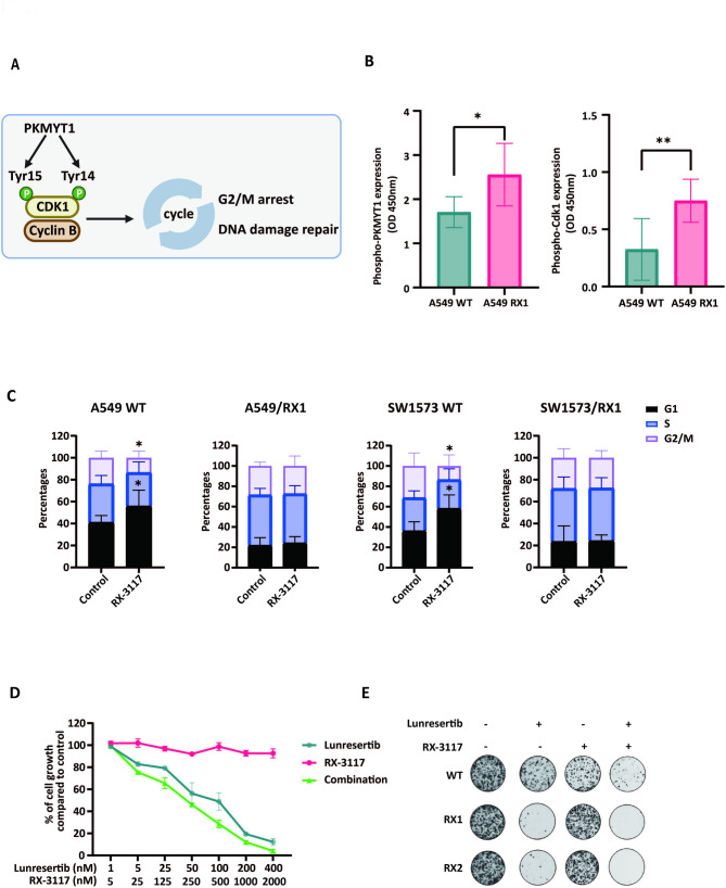
Results: Resistant clones exhibited decreased accumulation of RX-3117 nucleotides, which however, was not associated to reduced expression of activation enzymes (UCK2, UMPK, CMPK, NME1/NDPK, RR1 and RR2). Instead, increased expression was observed in certain DNA repair and deactivation enzymes (NT5C3) but pharmacological inhibition and silencing of the latter did not circumvent resistance. Remarkably, a comprehensive approach with CRISPR-Cas9 screen highlighted DNA-repair and cell cycle determinants as key sensitizing genes. XL-PCR and RNA-sequencing confirmed aberrations in DNA-repair and pathways involved in cell cycle regulation. Knock-out and pharmacological inhibition validated the role of PKMYT1, a protein kinase involved in G2/M transition and genomic stability. RX-3117-resistant A549 cells showed enhanced sensitivity to the PKMYT1 inhibitor lunresertib and its synergism with RX-3117, suggesting further studies, especially in patients with high PKMYT1 expression who have significantly shorter survival rates, as observed in public databases and validated in an internal cohort of NSCLC patients.

Conclusion: By integrating CRISPR-Cas9 with functional assays and transcriptomics, our study established a framework for decoding resistance mechanisms and highlights potential therapeutic strategies to enhance RX-3117 efficacy in NSCLC. We demonstrated for the first time that aberrant DNA repair and cell cycle dysregulation led resistance, identifying PKMYT1 as a promising target.

Keywords: Cell cycle distribution; Chemoresistance; DNA repair; Non-small cell lung cancer; Nucleoside analogs; PKMYT1.


Conflict of interest statement

Declarations. Ethics approval and consent to participate: The study on patients’ specimens was approved by the local ethics committees (Ethics Committee of Istituto Clinico Humanitas (Rozzano, Milan, Italy)– Ref-No. 165/17, Comitato Etico di Area Vasta Nord Ovest (CEAVNO) Regione Toscana– Protocol No. 31677, University Hospital Antwerp (Antwerp, Belgium) ethics committee– Protocol No. B300201316249 and Onze Lieve Vrouw Hospital (Aalst, Belgium) ethics committee– Protocol No. B300201317801) and conducted in accordance with principles stated in the Declaration of Helsinki. Written informed consent was obtained from all patients. Consent for publication: Not applicable. Competing interests: The authors declare no competing interests.

Figures

Fig. 1
Fig. 1
RX-3117 resistant NSCLC models and the role of nucleotide accumulation. (A) RX-3117 resistant cell lines were established by chronic exposure starting from A549 parental (wild type, WT), SW1573 WT, H460 WT cells and the gemcitabine resistant SW1573/G- cells. (B) Time course of exposure and stepwise increase in RX-3117 concentrations to produce resistant variants. (C, D) Growth inhibition curves of A549 (C) and SW1573 (D) resistant variants after 72-hr exposure to RX-3117. No growth inhibition was observed at the highest concentration of RX-3117 used, except for the SW/G/RX cell line. Results in the graph are from one representative experiment and bars represent the SD. IC50 values were calculated as mean of 3 independent experiments, each performed in triplicate. (E) Flow chart of the LC/MS-MS analysis. (F) Concentration dependent accumulation of RX-3117 nucleotides in A549 WT, SW1573 WT, SW1573/G- cells and depletion of RX-3117 nucleotides in RX-3117 resistant cells. Statistical significance was highlighted by asterisks, as follows: *P < 0.05, ***P < 0.001
Fig. 2
Fig. 2
Role of metabolism and DNA repair enzymes in RX-3117 acquired resistant cells and extent of accumulated DNA damage by XL-PCR. (A) Putative mechanism of action of RX-3117 and assumed components of the RX-3117 pathway that could contribute to resistance. (B) No common modulation of protein expression (i.e. increased or decreased expression) was observed for the resistant variants when considering the activation enzymes, UCK 2 clone 22 − 1 (UCK2 (22 − 1)), cytidine/uridine monophosphate kinase 1 (CMPK1), NME/NM23 nucleoside diphosphate kinase 1 (NME1/NDKA). A decreased expression of ribonucleotide reductase 1 and increased expression of ribonucleotide reductase 2 was observed in A549 cells (RR1 and RR2). Conversely, significantly increased expression of NT5C3 in both resistant variants was observed (representative blots, with 30 µg protein loading per sample, using β-actin as a loading control). (C) Increased expression of DCTPP1, NUDT1, TDP1, SAMHD1 was detected in both resistant variants, though more pronounced in RX1 cells (representative blots, with 40 µg protein loading per sample using β-actin as loading control). (D) Flow chart of the methodology used for the XL-PCR. (E) β-globin amplification as a measure of accumulated DNA damage was determined in A549 WT, SW1573 WT, SW1573/G- cells and RX-3117 resistant cells. Data represent the means ± SD from 3 individual experiments. Statistical significance was highlighted by asterisks, as follows: *P < 0.05, **P < 0.01, ***P < 0.001
Fig. 3
Fig. 3
CRISPR library screening and transcriptomics identified PKMYT1 and associated cell cycle and DNA repair genes pathways as drivers for RX-3117 resistance. (A) Graphical representation of the workflow for the CRISPR screen performed in A549 cells with TKO v3 whole-genome gRNA library. (B) Volcano plot showed the significant sensitizing and desensitizing genes as reflected by the indicated sensitizing and desensitizing gRNAs. The fold change (Log2) is plotted on the x-axis and the significance (− Log10 p-value) is plotted on the y-axis. The most significant sensitizing gene (PKMYT1) was highlighted. (C) Top 15 pathways enriched in sensitizing genes, as determined by the GSEA analysis (*P < 0.05). Red color indicates cell cycle pathway, and blue color indicates DNA repair pathway. (D) Top 15 pathways were enriched in the RNA-seq data between A549-WT and A549-RX1 cells, using the GSEA analysis, with red color indicating cell cycle pathway. (E) The GeneMANIA network showed that PKMYT1 is closely associated with key genes regulating the cell cycle
Fig. 4
Fig. 4
Validation of CRISPR Cas9 knockout status and the effect of cell cycle regulator PKMYT1 on RX-3117 sensitivity (A) Expression level of PKMYT1 by q-RT-PCR in A549 WT and single knock out clones KO1 and KO2. (B) Enzyme-Linked Immunosorbent Assay (ELISA) of phospho-PKMYT1 in A549 WT and KO1 and KO2 clones. (C, D) Colony formation assay performed on A549 WT and KO1 and KO2 clones, treated with different concentrations of RX-3117 in comparison with the negative control. Histogram bars show the means and SD of at least three experiments. Statistical significance was highlighted by asterisks, as follows: *P < 0.05; **P < 0.01
Fig. 5
Fig. 5
Differential perturbation of cell cycle induced by RX-3117 in RX-3117 resistant cells and inhibitory effects of lunresertib. (A) Graphical representation of the role of PKMYT1 on the regulation of cell cycle. (B) Enzyme-Linked Immunosorbent Assay (ELISA) of phospho-PKMYT1 and phospho-CDK1 in A549 WT and resistant model RX1. (C) Effect of RX3117 on the cell cycle distribution of A549 WT; A549 RX1; SW1573 WT; and SW1573 RX1 cells. (D) Growth inhibition curves of A549-RX1 resistant cells following exposure to lunresertib alone, RX3117 alone, or their combination. (E) Colony formation assay performed on A549-WT and RX1 cells treated with the 4X IC50 concentration of lunresertib and RX3117. Statistical significance was highlighted by asterisks, as follows: *P < 0.05; **P < 0.01
Fig. 6
Fig. 6
PKMYT1 expression and survival analysis in lung cancer patients. (A) Graphs reporting the mRNA expression levels of PKMYT1 in lung cancer tissues and normal tissues in TCGA-LUAD. (B) Kaplan–Meier analysis of overall survival in the patients of the TCGA-LUAD cohort (n = 502), stratified according to the high or low PKMYT1 mRNA expression levels. (C) Representative pictures of PKMYT1 protein staining in NSCLC tissues (left panel showing low expression, right panel showing high expression. Scale bars: 100 μm). (D) Kaplan–Meier analysis of overall survival in NSCLC patients (n = 66) stratified according to the high or low PKMYT1 protein expression levels. (E) Kaplan–Meier analysis of relapse-free survival in NSCLC patients (data on relapse were available for n = 45 patients, treated with gemcitabine-based regimens) stratified according to the high or low PKMYT1 protein expression levels

References

    1. Ciccolini J, Serdjebi C, Peters GJ, Giovannetti E. Pharmacokinetics and pharmacogenetics of gemcitabine as a mainstay in adult and pediatric oncology: an EORTC-PAMM perspective. Cancer Chemother Pharmacol. 2016;78(1):1–12. - PMC - PubMed
    1. Elnaggar M, Giovannetti E, Peters GJ. Molecular targets of gemcitabine action: rationale for development of novel drugs and drug combinations. Curr Pharm Des. 2012;18(19):2811–29. - PubMed
    1. Trial.gov C. Dose-Finding and Safety Study for Oral Single-Agent to Treat Advanced Malignancies: ClinicalTrials.gov; [Available from: https://www.clinicaltrials.gov/study/NCT02030067?intr=RX-3117&rank=2
    1. Salgia N, Pal SK, Chung V, Tagawa ST, Picus J, Babiker HM et al. Activity of RX-3117, an oral antimetabolite nucleoside, in subjects with advanced urothelial cancer: preliminary results of a phase IIa study. J Clin Oncol. 2019;37(7).
    1. Babiker H, Schlegel PJ, Hicks LG, Bullock AJ, Burhani N, Mahadevan D, et al. A multicenter phase 1/2 study investigating the safety, pharmacokinetics, pharmacodynamics and efficacy of a small molecule antimetabolite, RX-3117, plus nab-paclitaxel in pancreatic adenocarcinoma. Invest New Drugs. 2022;40(1):81–90. - PubMed

MeSH terms