Review
. 2025 Jul 9:16:1608612.
doi: 10.3389/fendo.2025.1608612. eCollection 2025.

Unraveling the role of STAT3 in Cancer Cachexia: pathogenic mechanisms and therapeutic opportunities

Affiliations
Review

Unraveling the role of STAT3 in Cancer Cachexia: pathogenic mechanisms and therapeutic opportunities

Xinyi Lv et al. Front Endocrinol (Lausanne). .

Abstract

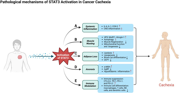
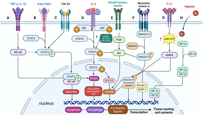
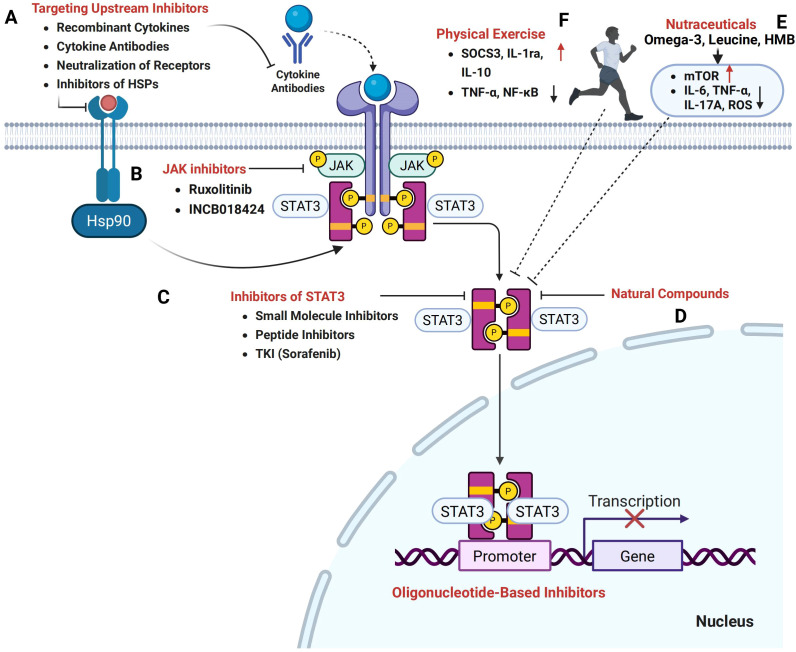
Cancer cachexia is a complex, multifactorial syndrome characterized by severe weight loss, muscle wasting, and systemic inflammation, significantly contributing to cancer-related morbidity and mortality. Signal transducer and activator of transcription 3 (STAT3) has emerged as a central mediator in the pathogenesis of this multifactorial condition. STAT3 regulates a broad range of cellular processes including inflammation, proteolysis, and mitochondrial dysfunction across multiple tissues, particularly skeletal muscle and adipose tissue. Persistent activation of STAT3 in response to tumor-derived and host-derived cytokines drives catabolic signaling cascades, disrupts anabolic pathways, and impairs energy homeostasis. Recent studies have illuminated the cross-talk between STAT3 and other signaling pathways that exacerbate cachexia-related metabolic imbalances. These findings position STAT3 not only as a critical mediator of cachexia progression but also as a promising therapeutic target. Pharmacological inhibition of STAT3 signaling has demonstrated efficacy in preclinical models, offering potential avenues for clinical intervention. This review provides a comprehensive overview of the molecular mechanisms by which STAT3 contributes to cancer cachexia and discusses emerging therapeutic strategies aimed at modulating STAT3 activity to mitigate the progression of this debilitating syndrome.

Keywords: STAT3; cancer cachexia; muscle wasting; pathogenic mechanisms; systemic inflammation; therapeutic strategies.


Conflict of interest statement

The authors declare that the research was conducted in the absence of any commercial or financial relationships that could be construed as a potential conflict of interest.

Figures

Figure 1
Figure 1
Activation of STAT3 signaling pathways and their role in cancer cachexia. (A) Both canonical and non-canonical STAT3 signaling pathways are crucial for cellular signaling. In the canonical pathway, cytokines such as IL-6, LIF, and OSM bind to their receptors, inducing receptor dimerization and the recruitment of JAKs. This interaction results in receptor phosphorylation at specific tyrosine residues, creating docking sites for STAT3. STAT3 is then phosphorylated by JAKs, dissociates from the receptor, forms homodimers or heterodimers, and translocates to the nucleus to regulate gene transcription. In contrast, the non-canonical STAT3 pathway involves mitochondrial STAT3 (mtSTAT3), unphosphorylated STAT3, and serine 727-phosphorylated STAT3 (p-STAT3 Ser727), alone or in combination with tyrosine 705-phosphorylated STAT3 (p-STAT3 Tyr705). These variants play a role in mitochondrial function, emphasizing STAT3’s involvement beyond transcriptional regulation. (B) In cancer cachexia, STAT3 activation occurs through both receptor- and non-receptor-mediated mechanisms. Pro-inflammatory cytokines or growth factors bind to cell surface receptors, leading to tyrosine phosphorylation, which facilitates the recruitment of JAKs or direct binding of STAT3 via its Src homology 2 (SH2) domain. Phosphorylated STAT3 dimerizes and enters the nucleus, promoting the transcription of genes involved in catabolic processes, driving tissue degradation and metabolic imbalance.
Figure 2
Figure 2
Pathological mechanisms of STAT3 in cancer cachexia. This figure illustrates the diverse roles of STAT3 activation in cancer patients, emphasizing its contribution to systemic inflammation, muscle atrophy, metabolic dysfunction, appetite regulation, and immune suppression. (A) In systemic inflammation, activated STAT3 triggers the release of pro-inflammatory cytokines such as IL-6, IL-1, and COX-2, exacerbates inflammation in the central nervous system, and contributes to anorexia. (B) In skeletal muscle, STAT3 activation induces atrophy by stimulating the ubiquitin–proteasome system and autophagy-related pathways, while impairing muscle regeneration by disrupting the differentiation, proliferation, and self-renewal of muscle satellite cells. Additionally, it compromises mitochondrial function. (C) In adipose tissue, activated STAT3 promotes lipolysis and metabolic dysregulation, inhibits lipogenesis, and suppresses brown adipose tissue differentiation and the expression of uncoupling protein 1 (UCP1). (D) In appetite regulation, STAT3 enhances the activity of pro-opiomelanocortin (POMC) neurons, increasing α-melanocyte-stimulating hormone (α-MSH) production to promote satiety, while simultaneously suppressing agouti-related peptide (AgRP) neurons that normally stimulate hunger, leading to reduced food intake and body weight loss. (E) STAT3 contributes to immune suppression by upregulating immune checkpoint molecules such as CTLA-4, PD-1, PD-L1, and PD-L2. Its activation alters the differentiation and function of immune cells, including macrophages, T cells, natural killer (NK) cells, and dendritic cells, reshaping the immune microenvironment and accelerating tumor progression.
Figure 3
Figure 3
Signaling cross-talk between STAT3 and other pathways in cancer cachexia. (A) IFNγ/TNFα signaling induces phosphorylation of STAT3 at Y705, promoting its interaction with NF-κB to form a nuclear complex that activates the iNOS/NO pathway, a critical mediator of muscle loss. (B) TNF-α and IL-1β upregulate SOCS-box protein 1 (SPSB1) expression through NF-κB signaling. (C) IL-6 enhances SPSB1 expression via the glycoprotein 130/JAK2/STAT3 pathway, while TGF-β activates STAT3 in a SMAD-dependent manner. (D) TGFβ1 induces Tyr705 phosphorylation of STAT3 in C2C12 cells. (E) STAT3 signaling interacts with the PI3K/Akt/mTOR pathway by suppressing p-Akt activity. (F) Myostatin and Activin A activate SMAD2/3 signaling similarly and inhibit the insulin/IGF-1/Akt/mTOR pathway, reducing muscle mass and function. (G) IL-6 increases AMPK activity in C2C12 cells and mouse cancer cachexia models, and AMPK activation enhances myofibrillar protein degradation. (H) HIF-1α shifts muscle metabolism by upregulating glycolysis and downregulating oxidative phosphorylation. (I) The role of miRNAs in regulating STAT3 activation and enhancing its cachectic effects.
Figure 4
Figure 4
Therapeutic potential of targeting the STAT3 signaling pathway in cancer cachexia. (A) Therapeutic strategies aimed at upstream inhibitors, including recombinant cytokines, cytokine antibodies, receptor neutralization, and inhibitors of heat shock proteins (HSPs), hold promise in modulating STAT3 activation and mitigating cachexia. (B) Janus kinase (JAK) inhibitors, such as Ruxolitinib and INCB018424, effectively inhibit the JAK/STAT3 pathway. (C) Direct STAT3 inhibition can be achieved through small molecule inhibitors, peptide inhibitors, and tyrosine kinase inhibitors (TKIs) such as Sorafenib, which prevent STAT3 dimerization and nuclear translocation. (D) Natural compounds, including plant-derived phytochemicals, also target the STAT3 signaling axis. (E) Nutritional and metabolic modulators, particularly nutraceuticals, have the potential to influence STAT3-mediated pathways and restore the balance between protein synthesis and degradation. (F) Furthermore, exercise interventions targeting STAT3 signaling offer a promising strategy to counteract the muscle wasting and metabolic imbalance characteristic of cancer cachexia.

Similar articles

References

    1. Setiawan T, Sari IN, Wijaya YT, Julianto NM, Muhammad JA, Lee H, et al. Cancer cachexia: molecular mechanisms and treatment strategies. J Hematol Oncol. (2023) 16:54. doi: 10.1186/s13045-023-01454-0, PMID: - DOI - PMC - PubMed
    1. Baracos VE, Martin L, Korc M, Guttridge DC, Fearon KCH. Cancer-associated cachexia. Nat Rev Dis Primers. (2018) 4:17105. doi: 10.1038/nrdp.2017.105, PMID: - DOI - PubMed
    1. Wu Q, Li B, Li J, Sun S, Yuan J, Sun S. Cancer-associated adipocytes as immunomodulators in cancer. biomark Res. (2021) 9:2. doi: 10.1186/s40364-020-00257-6, PMID: - DOI - PMC - PubMed
    1. Wu Q, Liu Z, Li B, Liu YE, Wang P. Immunoregulation in cancer-associated cachexia. J Adv Res. (2024) 58:45–62. doi: 10.1016/j.jare.2023.04.018, PMID: - DOI - PMC - PubMed
    1. Nishikawa H, Goto M, Fukunishi S, Asai A, Nishiguchi S, Higuchi K. Cancer cachexia: its mechanism and clinical significance. Int J Mol Sci. (2021) 22:8491. doi: 10.3390/ijms22168491, PMID: - DOI - PMC - PubMed

Substances

LinkOut - more resources