. 2025 Jul 4;5(4):oeaf083.
doi: 10.1093/ehjopen/oeaf083. eCollection 2025 Jul.

Atrial granules as acidic calcium stores in cardiomyocytes

Affiliations

Atrial granules as acidic calcium stores in cardiomyocytes

Emily Akerman et al. Eur Heart J Open. .

Abstract

Acidic calcium stores significantly influence basal calcium transient amplitude and β-adrenergic responses in cardiomyocytes. Atrial myocytes contain atrial granules (AGs), small acidic organelles that store and secrete atrial natriuretic peptide (ANP) and are absent in healthy ventricular myocytes. AGs are known to be acidic and calcium-rich, but their number and location relative to other signalling sites remain unexplored. Labelling of acidic organelles in adult guinea pig cardiomyocytes showed the presence of acidic puncta throughout the cytosol. Atrial myocytes exhibited an increased concentration of acidic organelles at the nuclear poles. Live cell fluorescent studies using 4-phenyl-3-butenoic acid (PBA) to inhibit peptidylglycine α-amidating monooxygenase, a crucial component of AGs membranes, effectively eliminated staining at the nuclear poles and most acidic puncta in atrial cells, but not in ventricular cells. Our immunofluorescent labelling also emphasizes the differences in acidic punctae between atrial and ventricular myocytes by showing minimal co-localization between AG-specific ANP and lysosomal-associated membrane protein. Electron microscopy studies on goat atrial fibrillation (AF) and sham control tissue allowed visualization of AGs. Quantitative analysis revealed that AGs were positioned significantly further away from the nearest sarcoplasmic reticulum and were closer to mitochondria in AF compared to sinus rhythm control tissue. We raise the question whether the positioning of AGs is strategic for communication with other calcium-containing organelles.

Keywords: Atrial fibrillation; Atrial granules; Cardiomyocytes; Electron microscopy; PAM.


Conflict of interest statement

Conflict of interest: None to declare.

Figures
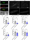
Figure 1
Figure 1
Atrial granules comprise the majority of acidic organelles in live atrial myocytes. (A) Representative example of a fixed, isolated guinea pig atrial myocyte immunolabelled for (i) atrial natriuretic peptide (green), (ii) lysosomal-associated membrane protein 2 (red), and (iii) co-immunolabelled for atrial natriuretic peptide and lysosomal-associated membrane protein 2; brightfield image is shown in (iv). (B) LysoTracker™ staining of acidic organelles (100 nM, 20 min) in primary isolated guinea pig atrial myocytes in control conditions (i) and in the presence of 4-phenyl-3-butenoic acid (200 µM, 2 h; ii) and in ventricular myocytes in control conditions (iii) and in the presence of 4-phenyl-3-butenoic acid (200 µM, 2 h; iv). (C) (i) Acidic perinuclear puncta count in control conditions (vehicle control n = 10) and in the presence of 4-phenyl-3-butenoic acid (200 µM, n = 14) in atrial myocytes; (ii) acidic peripheral puncta count in control conditions (vehicle control, n = 10) and 4-phenyl-3-butenoic acid (200 µM, n = 14) at the subsarcolemmal-apical region (top) of atrial myocytes; (iii) acidic peripheral puncta count in control conditions (vehicle control, n = 10) and 4-phenyl-3-butenoic acid (200 µM, n = 14) at the subsarcolemmal-basal region (bottom) of atrial myocytes. (D) (i) Acidic perinuclear puncta count in control conditions (vehicle control, n = 6) and 4-phenyl-3-butenoic acid (200 µM, n = 12) in ventricular myocytes; (ii) acidic peripheral puncta count in control conditions (vehicle control, n = 6) and 4-phenyl-3-butenoic acid (200 µM, n = 12) at the subsarcolemmal-apical region (top) of ventricular myocytes; (iii) acidic peripheral puncta count in control conditions (vehicle control, n = 6) and 4-phenyl-3-butenoic acid (200 µM, n = 12) at the subsarcolemmal-basal region (bottom) of ventricular myocytes. PBA, 4-phenyl-3-butenoic acid. Scale bar represents 20 μm.
Figure 2
Figure 2
Atrial granule position changes significantly in a goat model of atrial fibrillation. (A) Maximum atrial granule diameter in sham-operated (n = 35) and atrial fibrillation goat (n = 50). (B) Minimum distance between atrial granules and nearest sarcoplasmic reticulum measured in sham-operated (n = 34) and atrial fibrillation goat tissue (n = 46). (C) Minimum distance between atrial granules and mitochondria in sham-operated (n = 34) and atrial fibrillation goats (n = 48). (D) Summary table indicating maximum diameter, nearest sarcoplasmic reticulum, and nearest mitochondria to atrial granules measured in sham-operated and atrial fibrillation goat tomograms. AGs, atrial granules; SR, sarcoplasmic reticulum; AF, atrial fibrillation. (E) 3D tomography snapshot (i) and the indicated zoomed-in area (square; ii) from the left atrium goat sample in sinus rhythm control. (F) 3D tomography snapshot (i) and the indicated zoomed-in area (square; ii) from the left atrium goat sample in atrial fibrillation. Snap shots in (E) and (F) show proximity of atrial granules (AG) to mitochondria (Mito) and sarcoplasmic reticulum (SR arrows). Scale bars provided on each image represent 200 nm.

References

    1. Collins TP, Bayliss R, Churchill GC, Galione A, Terrar DA. NAADP influences excitation-contraction coupling by releasing calcium from lysosomes in atrial myocytes. Cell Calcium 2011;50:449–458. - PubMed
    1. Capel RA, Bolton EL, Lin WK, Aston D, Wang Y, Liu W, Wang X, Burton R-AB, Bloor-Young D, Shade K-T, Ruas M, Parrington J, Churchill GC, Lei M, Galione A, Terrar DA. Two-pore channels (TPC2s) and nicotinic acid adenine dinucleotide phosphate (NAADP) at lysosomal-sarcoplasmic reticular junctions contribute to acute and chronic β-adrenoceptor signaling in the heart. J Biol Chem 2015;290:30087–30098. - PMC - PubMed
    1. Aston D, Capel RA, Ford KL, Christian HC, Mirams GR, Rog-Zielinska EA, Kohl P, Galione A, Burton RAB, Terrar DA. High resolution structural evidence suggests the sarcoplasmic Reticulum forms microdomains with acidic stores (lysosomes) in the heart. Sci Rep 2017;7:40620. - PMC - PubMed
    1. Somlyo AV, Broderick R, Shuman H, Buhle EL, Somlyo AP. Atrial-specific granules in situ have high calcium content, are acidic, and maintain anion gradients. Proc Natl Acad Sci U S A 1988;85:6222–6226. - PMC - PubMed
    1. Cantin M, Genest J. The heart as an endocrine gland. Sci Am 1986;254:76–81. - PubMed

LinkOut - more resources