. 2025 Aug 15;138(16):jcs263736.
doi: 10.1242/jcs.263736. Epub 2025 Aug 15.

The mammalian protein MTCH1 can function as an insertase

Affiliations

The mammalian protein MTCH1 can function as an insertase

Anna Roza Dimogkioka et al. J Cell Sci. .

Abstract

The outer mitochondrial membrane (OMM) hosts a variety of proteins such as import machineries, enzymes, fission and fusion factors, and pore proteins. In Saccharomyces cerevisiae, the MIM complex, consisting of Mim1 and Mim2, mediates the insertion of α-helical proteins into the OMM. Until recently, it was unclear which proteins served this function in higher eukaryotes. Recent studies have identified MTCH2 as the insertase responsible for inserting α-helical proteins into the OMM in mammals. MTCH1 is a paralogue of MTCH2 but its general function and contribution to the biogenesis process are not clear. To better characterize MTCH1, we explored whether MTCH1 or MTCH2 could functionally replace Mim1 and/or Mim2 in yeast. Expression of MTCH1 and MTCH2 in yeast cells lacking Mim1, Mim2 or both revealed that MTCH1, but not MTCH2, could compensate for the growth defects upon deleting the MIM complex. Furthermore, MTCH1 could restore the biogenesis of MIM substrates, translocase of the outer membrane (TOM) complex stability and morphology of mitochondria. These findings indicate that MTCH1, by itself, has insertase activity and is a functional equivalent for the MIM complex, despite the absence of any evolutionary relation between the mammalian and yeast insertases.

Keywords: Insertase; MIM; MTCH1; MTCH2; Mitochondria; Outer membrane.


Conflict of interest statement

Competing interests The authors declare no competing or financial interests.

Figures
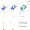
Fig. 1.
Fig. 1.
MTCH1 and MTCH2 are multi-span proteins that share structural similarity. Structural prediction of MTCH1 (UniProt: Q9NZJ7), MTCH2 (UniProt: Q9Y6C9), Mim1 (Uniprot: Q08176) and Mim2 (Uniprot: Q3E798) adapted from AlphaFold. Overlay of MTCH1 and MTCH2 structural predictions is also included. MTCH1 and MTCH2 structural predictions were aligned using Chimera X. Bottom right shows model confidence table corresponding to the AlphaFold structures. Protein structural models were computed using AlphaFold (https://alphafold.ebi.ac.uk/) using the amino acid sequences of the relevant proteins as input.
Fig. 2.
Fig. 2.
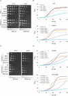
MTCH1 can complement the growth defects in cells lacking Mim1, Mim2 or both. (A,C) The growth of the indicated strains was monitored by drop dilution assay on solid synthetic medium containing either glucose (SD) or galactose (SGal) at either 30°C or 37°C. The strains were transformed with an empty vector (Ø) or vector encoding the indicated protein. Plates were incubated for 2 days at the indicated temperature before pictures were taken. (B,D) The growth of the indicated strains at 37°C in liquid glucose-containing medium was observed for 40 h using OD600 measurements. At the beginning of the measurements (time 0), the strains were diluted to an OD600 of 0.1. Data in this figure is representative of at least three repeats.
Fig. 3.
Fig. 3.
The N-terminal segment of MTCH1 contributes to its capacity to complement the absence of Mim1. (A) The growth of the indicated strains was monitored by drop dilution assay on solid synthetic medium containing either glucose (SD) or galactose (SGal) at either 30°C or 37°C. The strains were transformed with an empty vector (Ø) or vector encoding the indicated protein. Plates were incubated for a few days at the indicated temperature before pictures were taken. (B) The growth of the indicated strains at 37°C in liquid glucose-containing medium was monitored for 60 h using OD600 measurements. At the beginning of the measurements (time 0), the strains were diluted to an OD600 of 0.1. (C,D) Fractions corresponding to cytosol (cyt), ER and mitochondria (mit) were isolated from the indicated cells expressing the depicted HA-tagged proteins. The samples were analysed by SDS-PAGE and immunodecorated with the indicated antibodies. Hep1 is a mitochondrial protein; Erv2 is an ER protein; hexokinase (Hxk1) is a cytosolic marker. Data in this figure is representative of at least three repeats.
Fig. 4.
Fig. 4.
MTCH1 rescues the reduced steady state levels and assembly defects in cells deleted for Mim components. (A) Subcellular fractionation of WT, mim1Δ, mim2Δ and mim1/2ΔΔ strains expressing MTCH1-HA. Cytosol (cyt), ER and mitochondrial (mit) fractions were obtained using differential centrifugation steps. The samples were analysed by SDS-PAGE and immunodecorated with the indicated antibodies. Hep1 is a mitochondrial protein; Erv2 is an ER protein; hexokinase (Hxk1) is a cytosolic marker. (B) Mitochondria (100 µg) isolated from the indicated strains were analysed by SDS-PAGE and immunodecoration with antibodies against the indicated mitochondrial proteins. (C) The bands corresponding to the indicated proteins from three independent experiments as the one shown in B were quantified and normalized to the intensities of the Ponceau S staining. The value in the WT cells with empty plasmid was set as 100%. The bar diagram shows the mean±s.d. of three independent experiments. (D) Isolated mitochondria from the indicated strains were solubilized with 1% digitonin. Samples were analysed by BN-PAGE and immunodecorated with antibodies against Tom40. The bands representing the assembled TOM complex are indicated. Asterisk (*) indicates dissociated form of the TOM complex. (E) Radiolabelled pSu9-DHFR was synthesized in a cell-free system and imported into mitochondria isolated from the indicated cells. After import for the indicated times, proteinase K was added. Samples were analysed by SDS-PAGE and autoradiography. The precursor (p) and mature (m) forms of the protein are indicated. (F) Radiolabelled Tom20 was imported for the specified time periods into mitochondria isolated from the indicated cells. Samples were analysed via BN-PAGE and autoradiography. The band corresponding to the assembled TOM complex is indicated. Data in A, D–F is representative of three repeats.
Fig. 5.
Fig. 5.
MTCH1 can complement the mitochondrial morphology defect of mim1Δ cells. (A) mim1Δ cells harbouring mitochondria-targeted GFP (mtGFP) were transformed with either an empty plasmid (Ø) as a control (upper panels) or a plasmid encoding either MIM1 or MTCH1 (middle and lower panels, respectively). Cells were analysed by fluorescence microscopy and representative images of the predominant morphology for each strain are shown. Scale bar: 5 µm. (B) Quantification of the cells described in A where cells with either normal or abnormal mitochondrial morphology were counted. Mean±s.d. of three independent experiments with at least n=100 cells in each experiment are shown.

References

    1. Bartoš, L., Menon, A. K. and Vácha, R. (2024). Insertases scramble lipids: molecular simulations of MTCH2. Structure 32, 505-510.e4. 10.1016/j.str.2024.01.012 - DOI - PMC - PubMed
    1. Becker, T., Wenz, L. S., Krüger, V., Lehmann, W., Müller, J. M., Goroncy, L., Zufall, N., Lithgow, T., Guiard, B., Chacinska, A.et al. (2011). The mitochondrial import protein Mim1 promotes biogenesis of multispanning outer membrane proteins. J. Cell Biol. 194, 387-395. 10.1083/jcb.201102044 - DOI - PMC - PubMed
    1. Bhatia, S., Rawal, R., Sharma, P., Singh, T., Singh, M. and Singh, V. (2021). Mitochondrial dysfunction in Alzheimer's disease: opportunities for drug development. Curr. Neuropharmacol. 20, 675-692. 10.2174/1570159x19666210517114016 - DOI - PMC - PubMed
    1. Dimmer, K. S., Papić, D., Schumann, B., Sperl, D., Krumpe, K., Walther, D. M. and Rapaport, D. (2012). A crucial role for Mim2 in the biogenesis of mitochondrial outer membrane proteins. J. Cell Sci. 125, 3464-3473. 10.1242/jcs.103804 - DOI - PubMed
    1. Guna, A., Stevens, T. A., Inglis, A. J., Replogle, J. M., Esantsi, T. K., Muthukumar, G., Shaffer, K. C. L., Wang, M. L., Pogson, A. N., Jones, J. J.et al. (2022). MTCH2 is a mitochondrial outer membrane protein insertase. Science 378, 317-322. 10.1126/science.add1856 - DOI - PMC - PubMed

MeSH terms

Substances

LinkOut - more resources