. 2025 Nov 11;9(21):5436-5448.
doi: 10.1182/bloodadvances.2025016276.

HLA-E[pHLA-G] complex-specific monoclonal antibody enhancing NK activity in multiple myeloma

Affiliations

HLA-E[pHLA-G] complex-specific monoclonal antibody enhancing NK activity in multiple myeloma

Muhammad Abu Ahmad et al. Blood Adv. .

Abstract

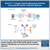
HLA-E presenting the HLA-G leader peptide VMAPRTLFL (HLA-E[pHLA-G]) on tumor cells plays a crucial role in suppressing natural killer (NK) and cytotoxic CD8+ T cells through NKG2A interaction. While blocking HLA-E:NKG2A is a promising immune checkpoint (IC) approach in cancer therapy, toxicity remains a major clinical concern. We developed a novel IC inhibitor that selectively prevents HLA-E:NKG2A interaction, a monoclonal antibody that selectively targets the HLA-E[pHLA-G] complex, distinguishing cancerous from noncancerous cells. In clinical bone marrow samples from patients with multiple myeloma (MM), 4D7 specifically recognized tumor-associated HLA-E-peptide complexes. Using NK cells from healthy donors, 4D7 effectively blocked the HLA-E:NKG2A interaction, and enhanced NKG2A-positive NK cell activity in autologous MM cell cocultures. Importantly, 4D7 did not inhibit NKG2C-positive NK cells, preserving their activity, even though NKG2C also interacts with HLA-E. In MM-bearing mice treated with human NK cells, 4D7 significantly reduced tumor growth. This targeted approach activates NK cells only against tumor cells presenting HLA-E-peptide complexes, potentially minimizing toxicity compared with current NKG2A inhibitors. The development of 4D7 highlights a promising advancement in immunotherapy for hematologic malignancies, offering improved outcomes for patients with MM, and a foundation for broader application across cancer types.


Conflict of interest statement

Conflict-of-interest disclosure: The authors declare no competing financial interests.

Figures
None
Graphical abstract
Figure 1.
Figure 1.
Recognition of recombinant HLA-E–peptide complexes by the mAb 4D7. (A) ELISA demonstrating the binding specificity of 4D7 or 3D12 to wells coated with 2 μg/mL of recombinant HLA-E[pHLAG] (blue bar), HLA-E [pHSP60] (light gray bar), or pHLA-G (dark gray bar). Murine IgG1κ isotype (2 μg/mL) served as the control. Absorption was measured at O.D. 650 nm. Error bars represent ± standard deviation (SD). Significance was tested by a 2-way analysis of variance (ANOVA). ∗∗∗∗P < .0001. (B) A ProteOn array showed the affinity of 4D7 at concentrations ranging from 0 to 40 nmol/L to 5 μg of HLA-E[pHLA-G]. Data were analyzed using the equilibrium model. (C-D) Binding model comparison of the variable chain of mAb 4D7 using ClusPro2 web server, depicted in blue and green (PDB ID: 3CDG), represented in shades of gray for the HLA-E alpha chain, and purple for the β2 microtubulin, in the presence (C) and absence (D) of the VMAPRTLFL peptide shown in red. O.D., optical density; W/o, without.
Figure 2.
Figure 2.
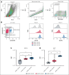
Staining of human leukemic cell lines for the HLA-E receptor and the acid-washing protocol for loading various peptides. (A) Staining of wild-type (WT) 721.221 and 721.221 HLA-G with mAb 3D12 (green panel) and 4D7 (blue panel). 0.2 × 10⁶ cells per well were incubated with the mAb PE-conjugated mouse anti-human HLA-E (3D12) and APC-conjugated mouse anti-human HLA-E (4D7) or a matched isotype control. (B) Schematic representation of the acid wash protocol, followed by flow cytometry determination of surface expression levels of HLA-E loaded with different nonapeptides. (C) HLA-E staining of acid-washed and peptide-loaded 721.221 HLA-G cells. 721.221 HLA-G cells were treated briefly with an acid buffer (citric acid in Na2HPO4 buffer, pH 5). Thirty-two different exogenous peptides, 20 μg/mL, with single amino acid mutations and the WT peptide were loaded on peptide-stripped 721.221 HLA-G cells. The treated cells were then stained with mAb 3D12 and mAb 4D7 or matched isotype control. The results were normalized to staining with commercial antibody to HLA-E. (D) Staining of U266, U937, and RPMI8226 cell lines with mAb 3D12 (green panel) and mAb 4D7 (blue panel). Murine IgG1k was used as an isotype control. For staining, all the antibodies were used at a concentration of 5 μg/mL. Samples were acquired by a CytoFLEX flow cytometer, and histograms were plotted using Kaluza software. PE, fluorescent dye R-phycoerythrin.
Figure 3.
Figure 3.
HLA-E expression in BMMCs from patients with MM. BMMCs were isolated from the BM of 6 patients with active MM who were treatment naïve. (A) Representative gating strategy for BM cell populations from patient 4. (B) Representative dot plot showing CD38 and CD138 subpopulations (left), and overlay histograms of HLA-E staining using mAb 3D12 (PE-conjugated, middle) and mAb 4D7 (APC-conjugated, right) in CD38CD138 (gray), CD38+CD138 (red), and CD38+CD138+ (blue) subpopulations. (C-D) Box plots summarizing HLA-E staining GMFI across the subpopulations for all 6 patients with MM using 3D12 (C) and 4D7 (D) antibodies. Cells (5 × 10⁵) were stained with BV780-conjugated anti-CD38 and fluorescein isothiocyanate-conjugated anti-CD138 antibodies (5 μg/mL each). Data were acquired using a CytoFLEX flow cytometer and analyzed with Kaluza software. FSC-A, forward scatter area; GMFI, geometric mean fluorescence intensity; ns, not significant; PE, fluorescent dye R-phycoerythrin; SSC-A, side scatter area.
Figure 4.
Figure 4.
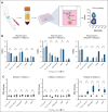
Impact of mAb 4D7 on primary human NK cell activity from healthy donors. (A) Schematic of the experimental design. Primary NK cells (5 × 10⁴) from healthy donors were cocultured with target cells (1.5 × 10⁵) for 4 hours in the presence of mAb 4D7 or matched isotype control (murine IgG1, 10 μg/mL). NK cell CD107a expression was then assessed by flow cytometry. (B) Representative CD107a degranulation percentage for donor 5. (C) Normalized mean results from 5 healthy donors. Panels show NK subsets: NKG2ANKG2C (top), NKG2ANKG2C+ (middle), and NKG2A+NKG2C (bottom). After incubation, NK cells were stained for subset markers (2 μg/mL). Data were acquired using a CytoFLEX flow cytometer and analyzed with GraphPad Prism v10. Isotype control degranulation was normalized to 1, with 4D7 mAb results adjusted accordingly. Experiments were performed in triplicate for each donor. Error bars represent ± SD. ∗P < .05; ∗∗∗∗P < .0001 (2-way ANOVA).
Figure 5.
Figure 5.
Impact of mAb 4D7 on MM-derived primary human NK cell activity. (A) Schematic of the experimental design. Primary NK cells were cocultured with target cell lines for 4 hours in the presence of 4D7 mAb or isotype control (murine IgG1, 10 μg/mL). (B) Normalized CD107a degranulation results for autologous (top), U266 (middle), and RPMI8226 (bottom) target cells. (C) Normalized IFN-γ production results for autologous (top), U266 (middle), and RPMI8226 (bottom) target cells. NK cells were cocultured with target cells (E:T ratio 1:3), and subsequently stained for CD107a, CD16, CD56, NKG2A, and NKG2C (2 μg/mL each). Supernatants were collected for IFN-γ measurements. Data were acquired using a CytoFLEX flow cytometer and analyzed with GraphPad Prism v10. CD107a-positive NK cell percentages were calculated and normalized as described in Figure 4. Results represent the average of 6 patients with MM, with experiments performed in triplicate for each donor. Error bars represent ± SD. ∗P < .05; ∗∗P < .01; ∗∗∗P < .001; ∗∗∗∗P < .0001 (2-way ANOVA). ns, not significant.
Figure 6.
Figure 6.
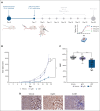
Evaluation of mAb 4D7 IC efficacy in a U266 CDX model implanted in NSG mice. (A) Experimental timeline illustrating U266 tumor implantation (day 0), pNK cell inoculation (day 7), hIL-15 supplementation and 4D7/control treatments from day 9, and tumor volume measurement schedule. (B) Tumor volume measurements of the different treatment groups, including vehicle, hIgG1, and mAb 4D7. (C) Ki67 staining quantification is presented as a boxplot. (D) Representative images of Ki67 staining in tumor sections from each treatment group. ∗∗P < .01; ∗∗∗∗P < .0001 (ANOVA). ns, not significant; pNK, primary NK cells.

References

    1. Kumar SK, Rajkumar V, Kyle RA, et al. Multiple myeloma. Nat Rev Dis Primers. 2017;3(1) - PubMed
    1. Siegel RL, Giaquinto AN, Jemal A. Cancer statistics, 2024. CA Cancer J Clin. 2024;74:12–49. - PubMed
    1. Bald T, Krummel MF, Smyth MJ, Barry KC. The NK cell–cancer cycle: advances and new challenges in NK cell–based immunotherapies. Nat Immunol. 2020;21(8):835–847. - PMC - PubMed
    1. Xu X, Zhou Y, Wei H. Roles of HLA-G in the maternal-fetal immune microenvironment. Front Immunol. 2020;11 - PMC - PubMed
    1. Yang Y, Wang W, Weng J, et al. Advances in the study of HLA class Ib in maternal-fetal immune tolerance. Front Immunol. 2022;13 - PMC - PubMed

MeSH terms

Substances