. 2025 Oct;116(10):2712-2722.
doi: 10.1111/cas.70144. Epub 2025 Jul 24.

Endothelial-Mesenchymal Transition in Tumor Microenvironment Promotes Neuroendocrine Differentiation of Prostate Cancer

Affiliations

Endothelial-Mesenchymal Transition in Tumor Microenvironment Promotes Neuroendocrine Differentiation of Prostate Cancer

Takumi Kageyama et al. Cancer Sci. 2025 Oct.

Abstract

Neuroendocrine prostate cancer (NEPC) is a highly aggressive and treatment-resistant subtype of castration-resistant prostate cancer (CRPC) that often emerges during progression under androgen-receptor (AR) pathway inhibition. While lineage plasticity in cancer cells has been recognized as a key mechanism of resistance, the role of the tumor microenvironment in driving this transition remains unclear. Among its cellular components, vascular endothelial cells can undergo endothelial-mesenchymal transition (EndoMT), a phenotypic shift associated with tumor progression and fibrosis. Here, we investigated whether EndoMT contributes to NEPC development. Human umbilical vein endothelial cells (HUVEC) were induced to undergo EndoMT using IL-1β and TGF-β2, and are hereafter referred to as EndoMTed HUVEC. EndoMTed HUVEC promoted neuroendocrine features and functional changes in LNCaP cells. Transcriptome analysis revealed marked upregulation of granulocyte-macrophage colony-stimulating factor (GM-CSF) in EndoMTed HUVEC. Neutralization of GM-CSF signaling using mavrilimumab, a monoclonal antibody targeting the GM-CSF receptor alpha (CSF2RA), and siRNA-mediated CSF2RA knockdown both suppressed the neuroendocrine phenotype and STAT3 signaling of LNCaP cells. Conversely, GM-CSF stimulation alone reproduced these changes. Enzalutamide-treated LNCaP cells secreted IL-1β and TGF-β2, which in turn triggered EndoMT, suggesting a reciprocal loop. These findings indicate that anti-androgen therapy may inadvertently promote NEPC through a paracrine loop involving tumor-derived cytokines and endothelial GM-CSF secretion, highlighting EndoMT as a microenvironmental driver of treatment resistance.

Keywords: androgen deprivation therapy; endothelial‐mesenchymal transition; granulocyte‐macrophage colony‐stimulating factor; neuroendocrine differentiation; prostate cancer.


Conflict of interest statement

Registry and the Registration No. of the Study/Trial: Not applicable.

Animal Studies: All animal experiments were approved by the Animal Research Committee of Mie University (approval number: 2019–32).

The authors declare no conflicts of interest.

Figures

FIGURE 1
FIGURE 1
Neuroendocrine differentiation‐like changes of LNCaP cells after co‐cultured with EndoMTed HUVEC. (A) Immunofluorescence staining of LNCaP cells co‐cultured with EndoMTed HUVEC (w/EndoMTed HUVEC), untreated HUVEC (w/HUVEC), or cultured alone, showing chromogranin A (CgA) expression. Scale bar: 200 μM. (B) CHGA mRNA expression in LNCaP cells under the same conditions, assessed by qPCR. (C) Western blotting of CgA and androgen receptor (AR) in LNCaP cells co‐cultured with EndoMTed HUVEC: β‐actin serves as a loading control. Representative of three independent experiments.
FIGURE 2
FIGURE 2
Phenotypical changes in androgen‐dependent prostate cancer cells, LNCaP, co‐cultured with EndoMTed HUVEC. (A) Brightfield microscope images of LNCaP cells alone, co‐cultured with untreated HUVEC (w/HUVEC), or with EndoMTed HUVEC (w/EndoMTed HUVEC) 24 h after seeding (start of co‐culture) and 96 h after co‐culturing. Scale bar 200 μM. (B) Cell proliferation rates at 96 h relative to 24 h under each condition. (C, D) Transwell migration assay using conditioned medium (CM) from HUVEC and EndoMTed HUVEC. Migrated cells were counted in five random fields per insert (20×). The average number of migrated cells per field was calculated. *p ≤ 0.05. (E) Heatmap of the cDNA microarray expression profiles in LNCaP cells under the same three conditions. (F) Western blotting of AR, CgA, FOXA2, FOXA1, and PSA. β‐actin was used as a loading control. Representative image shown. (G) Western blotting and qPCR of PSA and CHGA following 96 h co‐culture with EndoMTed HUVEC, and recovery culture in normal (androgen‐containing) medium. The medium was refreshed once at 48 h. AR, androgen receptor; CgA, chromogranin A; FOXA1, forkhead box A1; FOXA2, forkhead box A2; PSA, prostate‐specific antigen.
FIGURE 3
FIGURE 3
Expression analysis between HUVEC and EndoMTed HUVEC. (A) Heatmap of RNA‐seq analysis showing upregulated genes in EndoMTed HUVEC (n = 3). (B) Scatter plot highlighting differentially expressed genes (p‐value < 0.05, FDR < 0.01, Fold Change > 1.5). FDR, False Discovery Rate.
FIGURE 4
FIGURE 4
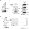
GM‐CSF–STAT3–MYC axis mediates neuroendocrine differentiation in LNCaP cells. (A) Western blotting of STAT3/MYC signaling pathway in LNCaP cells co‐cultured with HUVEC (w/HUVEC) or EndoMTed HUVEC (w/EndoMTed HUVEC). (B) Western blotting of NE markers and STAT3–MYC (c‐MYC) signaling in LNCaP cells co‐cultured with EndoMTed HUVEC and treated with mavrilimumab. (C) Western blotting of NE markers and STAT3–MYC signaling after siRNA‐mediated CSF2RA knockdown in LNCaP cells. (D) Western blotting of NE markers in LNCaP cells stimulated with recombinant GM‐CSF under androgen‐deprived conditions. (E) Transwell migration assay of LNCaP culture stimulated with GM‐CSF under androgen‐deprived conditions. (F) Quantification of migrated LNCaP cells; five random fields per insert were counted under a light microscope at 20× magnification. *p ≤ 0.05. CgA, chromogranin A; CSF2RA, colony‐stimulating factor 2 receptor alpha subunit; GM‐CSF, granulocyte‐macrophage colony‐stimulating factor; NE, neuroendocrine.
FIGURE 5
FIGURE 5
Elevated secretion of IL‐1β and TGF‐β2 in LNCaP cells, and EndoMT induction of HUVEC by LNCaP conditioned medium. (A) ELISA of IL‐1β and TGF‐β2 in culture media from LNCaP cells treated with or without enzalutamide (5 nM for 96 h, Selleck Chemicals, S1250). (B) Immunofluorescence staining of endothelial marker CD31 and mesenchymal marker αSMA in HUVEC exposed to conditioned media from untreated or enzalutamide‐treated LNCaP cells for 96 h, with one medium change at 48 h. *p ≤ 0.05. CD31, platelet endothelial cell adhesion molecule‐1; αSMA, alpha smooth muscle actin.
FIGURE 6
FIGURE 6
Histopathological features of human castration‐resistant prostate cancer tissues. Representative images of hematoxylin and eosin (HE) and immunofluorescence (IF) staining from three castration‐resistant prostate cancer (CRPC) cases with neuroendocrine differentiation (A–C). Colocalization analysis using ImageJ software quantitatively assessed the spatial relationship between vascular endothelial cells (CD31‐positive) and neuroendocrine cells (CgA‐positive), with Pearson's correlation coefficients (PCCs) of 0.39, 0.53, and 0.40 for patients A, B, and C, respectively (average PCC: 0.47). Green: CgA; Red: CD31; Blue: Nuclei. Scale bar: 50 μm. Clinical characteristics of each patient are summarized in Table S2. CgA, chromogranin A; CD31, platelet endothelial cell adhesion molecule‐1 (PECAM‐1).
FIGURE 7
FIGURE 7
LNCaP‐derived IL‐1β and TGF‐β2 induce HUVEC endothelial‐mesenchymal transition under androgen deprivation conditions, and the EndoMTed HUVEC, in turn, promotes LNCaP castration resistance.

References

    1. Maman S. and Witz I. P., “A History of Exploring Cancer in Context,” Nature Reviews Cancer 18, no. 6 (2018): 359–376. - PubMed
    1. Quail D. F. and Joyce J. A., “Microenvironmental Regulation of Tumor Progression and Metastasis,” Nature Medicine 19, no. 11 (2013): 1423–1437. - PMC - PubMed
    1. Kovacic J. C., Dimmeler S., Harvey R. P., et al., “Endothelial to Mesenchymal Transition in Cardiovascular Disease: JACC State‐Of‐The‐Art Review,” Journal of the American College of Cardiology 73, no. 2 (2019): 190–209. - PMC - PubMed
    1. Zeisberg E. M., Potenta S., Xie L., Zeisberg M., and Kalluri R., “Discovery of Endothelial to Mesenchymal Transition as a Source for Carcinoma‐Associated Fibroblasts,” Cancer Research 67, no. 21 (2007): 10123–10128. - PubMed
    1. Yoshimatsu Y., Wakabayashi I., Kimuro S., et al., “TNF‐α Enhances TGF‐β‐Induced Endothelial‐To‐Mesenchymal Transition via TGF‐β Signal Augmentation,” Cancer Science 111, no. 7 (2020): 2385–2399. - PMC - PubMed

MeSH terms