. 2025 Sep:219:107877.
doi: 10.1016/j.phrs.2025.107877. Epub 2025 Jul 23.

Combined dietary omega-3 and omega-6 fatty acids protect against hyperglycemia-associated retinopathy in neonatal mice

Affiliations

Combined dietary omega-3 and omega-6 fatty acids protect against hyperglycemia-associated retinopathy in neonatal mice

Myriam Boeck et al. Pharmacol Res. 2025 Sep.

Abstract

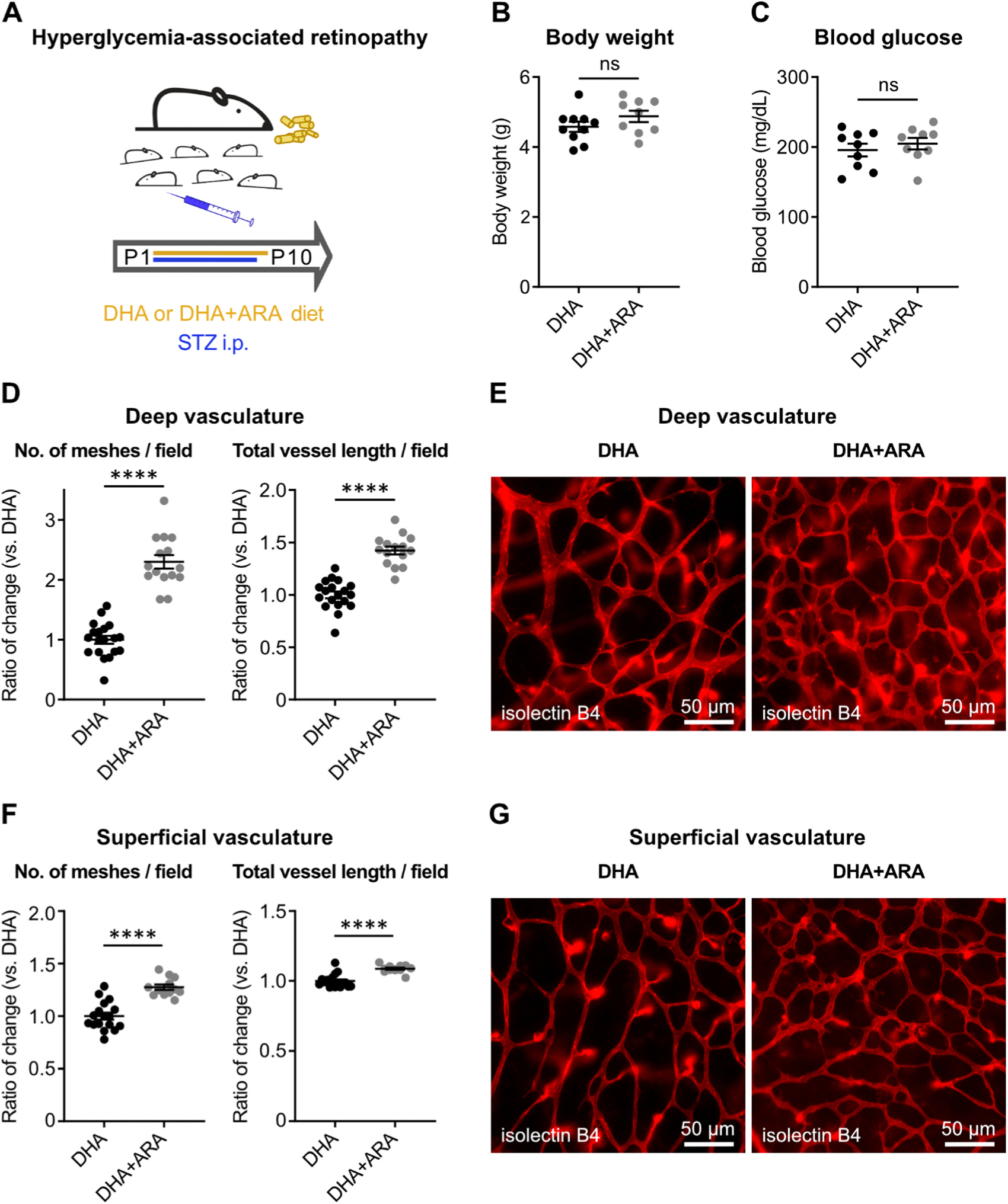
Retinopathy of prematurity (ROP) with early vessel loss (Phase I) followed by uncontrolled vessel growth (Phase II) causes visual impairment in premature infants. Although supplementation with omega-3 (n-3) docosahexaenoic acid (DHA) alone shows mixed results in preventing ROP, supplementation with both n-3 DHA and n-6 arachidonic acid (ARA) in early postnatal life reduces severe ROP by 50 % (Mega Donna Mega study). In the Mega Donna Mega study, 146 (72.6 %) of 201 included infants had at least one hyperglycemic episode during the first 14 days of life, which is a strong ROP risk factor. We therefore evaluated the protective effects and mechanisms of combined dietary n-3 DHA and n-6 ARA in a neonatal mouse model of hyperglycemia-induced suppression of retinal vascular development (Phase I ROP). At postnatal day (P) 10, retinal vessel growth was improved in pups from mothers on diets enriched with 1 % DHA + 2 % ARA vs. 3 % DHA. Lipid changes in pup plasma and RPE complex (retinal pigment epithelium with choroid and sclera) were in accordance with maternal diets' DHA and ARA levels, indicating that milk lipids reflected maternal diets. Proteomic retinal analysis revealed increased abundances of proteins related to mitochondrial respiration and glucose metabolism with the combined diet. Inhibition of mitochondrial ATP synthase negated the protective effects of the combined diet. In conclusion, combined DHA+ARA oral maternal supplementation protects against hyperglycemia-induced retinopathy in mouse neonates (Phase I ROP model) through enhanced retinal metabolism, suggesting the potential of balanced lipid supplementation for ROP prevention.

Keywords: ARA; DHA; Postnatal hyperglycemia; Retinal vasculature; Retinopathy of prematurity.


Conflict of interest statement

Declaration of Competing Interest The authors have declared no conflict of interest.

Figures

Fig. 1.
Fig. 1.
DHA+ARA diet vs. DHA promoted physiological retinal vascularization in hyperglycemia-associated retinopathy in neonatal mice. (A) Schematic of mouse model. To induce hyperglycemia-associated retinopathy, mouse pups were injected daily with streptozotocin (STZ, 50 μg/g body weight intraperitoneally) in PBS from postnatal day (P) 1–P9. Nursing moms were fed a diet supplemented with docosahexaenoic acid alone (DHA) or DHA plus arachidonic acid (DHA+ARA) from P1–P10. (B) P10 mouse pup body weight and (C) blood glucose levels were measured. DHA pups, n = 9–10; DHA+ARA pups, n = 9; unpaired t-test; ns, not significant. (D–G) Retinal vessel analysis of STZ-induced pups at P10. (D, F) Number of vascular meshes (left) and total vessel length (right) per field in deep (D) and superficial (F) retinal vasculature were quantified. DHA pup retinas, n = 17–19; DHA+ARA pup retinas, n = 12–15; unpaired t-test; **** p < 0.0001. (E, G) Representative images of deep (E) and superficial (G) retinal vasculature. Retinal vessels were visualized with isolectin B4 (red). Magnification, 200x; scale, 50 μm.
Fig. 2.
Fig. 2.
Maternal diet composition was reflected in the pup lipid profile of plasma and RPE complex. Targeted fatty acid analysis of neonatal mice with hyperglycemia-associated retinopathy. P10 (A) plasma, (B) RPE complex (retinal pigment epithelium with choroid and sclera), and (C) retina were isolated from mouse pups. Nursing moms were fed a diet supplemented with DHA or DHA+ARA from P1–P10. DHA pups, n = 18 (2 retinas pooled); DHA+ARA pups, n = 18 (2 retinas pooled); multiple unpaired t-test; * p < 0.05. Only comparisons with statistically significant changes are labeled. Fatty acids on the x-axis are denoted as Cx: yn-z, with x, number of carbons; y, number of double bonds; z, position of the first double bond from the omega (methyl) end.
Fig. 3.
Fig. 3.
DHA+ARA vs. DHA diet increased mitochondrial proteins in P10 retinas from mouse pups induced with hyperglycemia-associated retinopathy. Nursing moms were fed a diet supplemented with DHA or DHA+ARA from P1–P10. DHA pups, n = 8 (2 retinas pooled); DHA+ARA pups, n = 8 (2 retinas pooled). At P10, mouse retinas were isolated for proteomic analysis. (A) Principal component analysis of all detected proteins (≥ 2 unique peptides) in retinas isolated from mouse pups in DHA+ARA vs. DHA diet group. (B) Volcano plot of proteins with significantly (p < 0.05; q < 0.37; log2 fold change (log2FC) > 0.2 or < −0.2) increased (orange) or decreased (blue) abundance in retinas isolated from pups in DHA+ARA vs. DHA diet group. (C) Selected enriched (p < 0.05) gene ontology pathways associated with energy and lipid metabolism (biological process, BP; cellular component, CC; molecular function, MF) of proteins with significantly increased abundance (p < 0.05; log2FC > 0.2) in retinas isolated from pups in DHA+ARA vs. DHA diet group. Pathways related to oxidative phosphorylation are written in bold. Protein count refers to the number of distinct proteins with significantly increased abundance in the respective gene ontology cluster. Protein ratio describes the proportion of proteins with significantly increased abundance to all listed proteins in a cluster. (D) Heatmap depicting the altered proteins in the selected enriched pathways shown in (C).
Fig. 4.
Fig. 4.
Pharmacological inhibition of mitochondrial ATP production negated the promotion of physiological retinal vascularization by dietary DHA+ARA. (A) Schematic of intervention. Mouse pups induced with hyperglycemia-associated retinopathy were treated with mitochondrial ATP synthase inhibitor oligomycin A (25 μg/g body weight intraperitoneally) in 30 % dimethylsulfoxide in PBS from P7–P9. Nursing moms were fed DHA or DHA+ARA diet from P1–P10. (B) Body weight and (C) blood glucose levels of P10 oligomycin A-treated pups with maternal DHA or DHA+ARA diet were measured. DHA pups, n = 5–6; DHA+ARA pups, n = 8; unpaired t-test; ns, not significant. (D–G) Retinal vessel analysis of STZ-induced pups at P10. (D, F) Number of vascular meshes (left) and total vessel length (right) per field in deep (D) and superficial (F) retinal vasculature were quantified. DHA pup retinas, n = 12; DHA+ARA pup retinas, n = 16; unpaired t-test or Mann-Whitney U test; ns, not significant. (E, G) Representative images of deep (E) and superficial (G) retinal vasculature. Retinal vessels were visualized with isolectin B4 (red). Magnification, 200x; scale, 50 μm.

References

    1. Garcia H, Villasis-Keever MA, Zavala-Vargas G, Bravo-Ortiz JC, Perez-Mendez A, Escamilla-Nunez A, Global prevalence and severity of retinopathy of prematurity over the last four decades (1985–2021): a systematic review and meta-analysis, Arch. Med Res 55 (2) (2024) 102967. - PubMed
    1. Dai C, Xiao J, Wang C, Li W, Su G, Neurovascular abnormalities in retinopathy of prematurity and emerging therapies, J. Mol. Med. 100 (6) (2022) 817–828. - PMC - PubMed
    1. de Las Rivas Ramirez N, Luque Aranda G, Rius Diaz F, Perez Frias FJ, Sanchez Tamayo T, Risk factors associated with Retinopathy of Prematurity development and progression, Sci. Rep. 12 (1) (2022) 21977. - PMC - PubMed
    1. Tomita Y, Usui-Ouchi A, Nilsson AK, Yang J, Ko M, Hellstrom A, et al. , Metabolism in Retinopathy of Prematurity, Life (Basel) 11 (11) (2021). - PMC - PubMed
    1. Fu Z, Nilsson AK, Hellstrom A, Smith LEH, Retinopathy of prematurity: metabolic risk factors, Elife 11 (2022). - PMC - PubMed

MeSH terms