. 2025 Jul 24;16(1):6824.
doi: 10.1038/s41467-025-61864-1.

Restriction of Ku translocation protects telomere ends

Affiliations

Restriction of Ku translocation protects telomere ends

Stefano Mattarocci et al. Nat Commun. .

Abstract

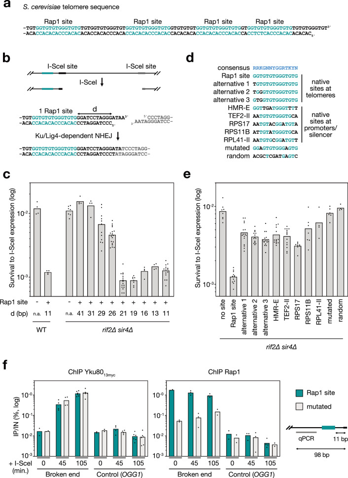
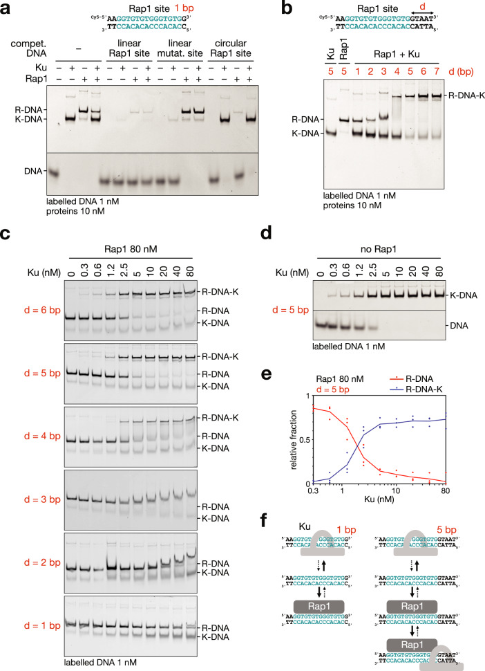
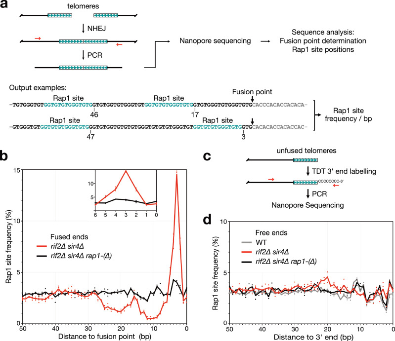
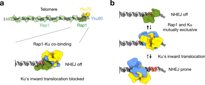
Safeguarding chromosome ends against fusions via nonhomologous end joining (NHEJ) is essential for genome integrity. Paradoxically, the conserved NHEJ core factor Ku binds telomere ends. How it is prevented from promoting NHEJ remains unclear, as does the mechanism that allows Ku to coexist with telomere-protective DNA binding proteins, Rap1 in Saccharomyces cerevisiae. Here, we find that Rap1 directly inhibits Ku's NHEJ function at telomeres. A single Rap1 molecule near a double-stand break suppresses NHEJ without displacing Ku in cells. Furthermore, Rap1 and Ku form a complex on short DNA duplexes in vitro. Cryo-EM shows Rap1 blocks Ku's inward translocation on DNA - an essential step for NHEJ at DSBs. Nanopore sequencing of telomere fusions confirms this mechanism protects native telomere ends. These findings uncover a telomere protection mechanism where Rap1 restricts Ku's inward translocation. This switches Ku from a repair-promoting to a protective role preventing NHEJ at telomeres.


Conflict of interest statement

Competing interests: The authors declare no competing interests.

Figures

Fig. 1
Fig. 1. Inhibition of NHEJ by a single Rap1 site at a DSB.
a Rap1 binding site at telomeres: Example of an S. cerevisiae telomere sequence (Rap1 sites in teal). b I-SceI NHEJ assay. Two inverted I-SceI sites were inserted at the URA3 locus. Survivors to continuous I-SceI expression eliminate fuse distal ends,. A Rap1 site was inserted near one I-SceI site. c NHEJ efficiency in wild-type (WT) and rif2∆ sir4∆ mutant cells with or without a Rap1 site at the break. Means from replicates, details and statistical analysis in Supplementary Table 1. d Distance between Rap1 and the break, (-) no insert, ( + ) 1 Rap1 site. d Sequences of the tested sites. Consensus from ref. (R: G/A, K: G/T, Y: T/C, N: any base). Bases identical to the telomeric Rap1 site (teal); differing bases (black). Alternative sites from native telomere sequences (Supplementary Fig. 4). Other sites from HMR-E silencer and TEF2, RPS17, RPS11 and RPL14 promoters. Negative controls: a mutated non-binding site and a 14-bp random sequence. e Divergent Rap1 sites from telomeres are less effective at blocking NHEJ. Sites 11 bp from the break. Cells lacking Rif2 and Sir4. Means from replicates, details and statistical analysis in Supplementary Table 1. f ChIP analysis of Yku80 and Rap1 binding at a DSB following 45 and 105 min of I-SceI induction. Data compare a Rap1 site (5GGTGTGTGGGTGTG3, teal) to a mutated site (5GGAGTGTGGGAGTG3, light grey). Cells were arrested in G1 to limit end resection. Quantification represents immunoprecipitated DNA (IP) relative to the input DNA (IN). Means from 2 independent cell cultures or more. OGG1 was used as a control locus. Source data are provided as a Source Data file.
Fig. 2
Fig. 2. Ku binding on Rap1-DNA complexes.
a Binding of Ku and Rap1 to a short DNA duplex containing a Rap1 site and a 1 bp downstream extension (1 nM). Unlabelled competitor DNA: (i) short linear duplex with a Rap1 site and a 1 bp downstream extension (titrates Rap1 and Ku, 200 nM), (ii) short linear duplex with a mutated site (mutated in Fig. 1) and a 1 bp downstream extension (titrates Ku only, 200 nM), circular plasmid with 16 Rap1 sites in tandem (titrates Rap1 only, 20 nM). (R) represents Rap1, (K) represents Ku. The experiment repeated three times. b Binding of Ku and Rap1 to DNA duplexes with downstream extensions ranging from 1 to 7 bp (1 nM). d refers number of base pairs separating the edge of the Rap1 site from the duplex end. The experiment repeated more than three times. c Representative titration of Ku binding to DNA duplexes with downstream extensions of 1 to 6 bp (1 nM) in the presence of excess Rap1 at a fixed concentration of 80 nM. d Representative titration of Ku binding to DNA in the absence of Rap1. The experiment repeated twice or more. e Quantified EMSA data showing Ku association on Rap1-bound DNA with a downstream extension of 5 bp (1 nM), as in (C). Means from 3 independent experiments. f Interpretative schematic illustrating the mutual exclusion or co-binding of Rap1 and Ku on short duplex DNA with a 1 or 5 bp extension downstream of the Rap1 site. Source data are provided as a Source Data file.
Fig. 3
Fig. 3. Structural analysis of Rap1’s ability to restrict Ku’s inward translocation on DNA.
Cryo-EM maps and corresponding models in cartoon representation of the Rap1DBD-DNA-Ku ternary complex (a, b) and the DNA-Ku binary complex (c, d). Yku70 is shown in gold, Yku80 in cornflower blue, Rap1DBD in green, the G-rich DNA strand in light grey and the C-rich DNA strand in grey. Nucleotides within 4.0 Å of Ku are highlighted in red on both strands. a, c The cryo-EM maps are shown in mesh and coloured according to atom proximity in the refined structural model. At the thresholds used, electron density was missing for residues 194–211 of Yku70, 165–171, 287–301, 462–466 of Yku80 and residues 429–433, 480–506 and 598–601 of Rap1DBD in the Rap1DBD-DNA-Ku complex (a) and for residues 200–211 of Yku70, 95–104, 165–171, 263–270, 286–300 and 579–587 of Yku80 in the DNA-Ku complex (c).
Fig. 4
Fig. 4. End-proximal Rap1 site position impacts telomere end protection.
a Schematic of the approach used to determine Rap1 site positions at fused telomeres. Two examples of sequenced telomere-telomere fusions are shown. Rap1 sites are highlighted in teal. b Rap1 site frequency at fused telomere ends. G-rich strand sequences. Fusion points at position 0. Means from independent cell cultures and sequencing experiments. c Schematic of the approach used to determine Rap1 site positions at unfused telomeres. X and Y’ telomere 3’ ends are labelled by TDT prior to PCR amplification. d Rap1 site frequency at unfused telomere ends. G-rich strand sequences. Telomere 3’ ends at position 0. Means from independent cell cultures and sequencing experiments. Source data are provided as a Source Data file.
Fig. 5
Fig. 5. Proposed model for telomere protection by the end-proximal Rap1.
a At telomeres where the end-proximal Rap1 site is not too close from the telomere double-stranded end, the restriction of Ku’s inward translocation by Rap1 protects telomeres. Yku70 is shown in gold, Yku80 in cornflower blue, end-proximal telomere-bound Rap1DBD in green, other telomere-bound Rap1DBD in light green, telomere DNA in grey, subtelomere DNA in light grey, with the Rap1 site highlighted in salmon (both strands). For simplicity, other telomere components, such as NHEJ inhibitors Rif2 and Sir4, are not represented. b At telomeres where the end-proximal Rap1 site is too close from the telomere double-stranded end to accommodate Ku when Rap1 is present, Rap1-Ku mutual exclusion predominates (same colour code as in panel a).

References

    1. de Lange, T. Shelterin-mediated telomere protection. Annu. Rev. Genet.52, 223–247 (2018). - PubMed
    1. Ruis, P. & Boulton, S. J. The end protection problem—an unexpected twist in the tail. Genes Dev.35, 1–21 (2021). - PMC - PubMed
    1. Chan, S. W.-L. & Blackburn, E. H. Telomerase and ATM/Tel1p protect telomeres from nonhomologous end joining. Mol. Cell11, 1379–1387 (2003). - PubMed
    1. Pobiega, S., Alibert, O. & Marcand, S. A new assay capturing chromosome fusions shows a protection trade-off at telomeres and NHEJ vulnerability to low-density ionizing radiation. Nucleic Acids Res.49, 6817–6831 (2021). - PMC - PubMed
    1. Stuart, A. & De Lange, T. Replicative senescence is ATM driven, reversible, and accelerated by hyperactivation of ATM at normoxia. Preprint at 10.1101/2024.06.24.600514 (2024).

MeSH terms

LinkOut - more resources