. 2025 Jul 24;15(1):26933.
doi: 10.1038/s41598-025-11990-z.

A hexamerization-enhanced, Fc-silenced agonistic CD27 antibody amplifies T-cell effector functions as single agent and in combination with PD-1 blockade

Affiliations

A hexamerization-enhanced, Fc-silenced agonistic CD27 antibody amplifies T-cell effector functions as single agent and in combination with PD-1 blockade

Işıl Altıntaş et al. Sci Rep. .

Abstract

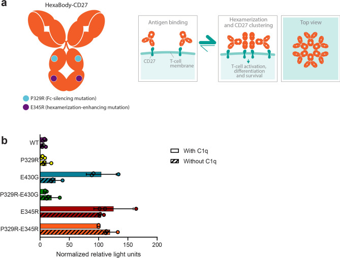
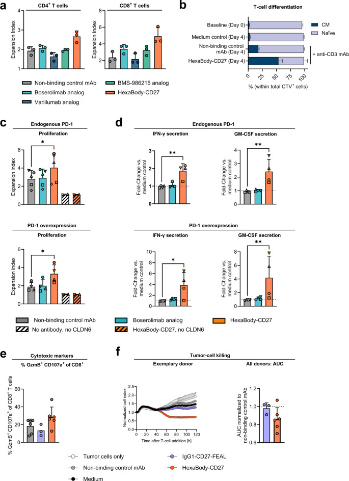
HexaBody-CD27 (GEN1053/BNT313) is an investigational novel agonistic CD27 antibody engineered to enhance T-cell costimulation and promote antitumor immunity. Through the introduction of a hexamerization-enhancing mutation in the IgG Fc domain, HexaBody-CD27 was designed to drive clustering and activation of CD27 via intermolecular Fc:Fc interactions between membrane-bound antibodies, independent of crosslinking by FcγR-bearing cells. HexaBody-CD27 carries an Fc-silencing mutation to prevent T-cell depletion through Fc-mediated effector functions. In vitro, HexaBody-CD27 induced CD27 receptor signaling independent of FcγR-mediated crosslinking in a reporter assay. It also enhanced T-cell proliferation, cytotoxic activity and proinflammatory cytokine secretion in primary human lymphocytes. In contrast to benchmark IgG1 CD27 antibodies, HexaBody-CD27 did not induce phagocytosis of T cells in vitro. HexaBody-CD27 promoted ex vivo tumor infiltrating lymphocyte (TIL) expansion in non-small cell lung cancer (NSCLC) specimens, in particular of CD8+ TILs. The combination of HexaBody-CD27 with an anti-PD-1 antibody enhanced T-cell proliferation, cytokine secretion, and cytotoxic activity in vitro compared to either compound alone. In conclusion, HexaBody-CD27 enhanced T-cell activation and effector functions in an FcγR-crosslinking-independent manner, without inducing T-cell depletion. The immune agonist activity of HexaBody-CD27 was potentiated in combination with PD-1 blockade.

Keywords: Combination therapy; Costimulatory molecules; Immunotherapy; Monoclonal antibody; T cell.


Conflict of interest statement

Declarations. Competing interests: UŞ and ÖT are management board members and employees at BioNTech (Mainz, Germany). KBN, AI, A-LK, AT, AM, FG, and SF-K are current or previous employees at BioNTech. Some of the authors have securities from BioNTech. IA, MMB, AI-F, JB, FJB, RNJ, DPES, BJK, MK, TA, and ECWB are current or previous employees at Genmab and own stock and/or stock options. IA, KBN, AIF, JB, AM, FJB, RNJ, DPES, FG, BJK, SF-K, ECWB, and UŞ are inventors on patents and/or patent applications related to CD27 HexaBody molecules.

Figures

Fig. 1
Fig. 1
A CD27 antibody with an Fc-silencing and a hexamerization-enhancing mutation drives CD27 agonist activity. (a) Proposed mechanism of action of HexaBody-CD27. Binding of HexaBody-CD27 to CD27 on T cells is hypothesized to result in the formation of hexameric antibody structures through Fc-Fc interactions between membrane-bound antibodies. Hexamer formation drives clustering and activation of CD27 receptors, resulting in enhanced T cell activation, differentiation and proliferation through CD27 agonism. (b) CD27 signaling in CD27-positive Jurkat reporter cells, after incubation with 10 µg/mL anti-CD27 antibody variants with a wild-type Fc (IgG1-CD27) or one or two Fc mutations: a P329R Fc-silencing mutation and/or an E430G or E345R hexamerization-enhancing mutation, in the absence or presence of 60 µg/mL purified C1q. Data shown are mean relative light units (RLU) +/− SD of three independent experiments, normalized by subtraction of non-binding control RLU values at 10 µg/mL (0%) and relative to 10 µg/mL of HexaBody-CD27 (P329R-E345R, 100%) in the presence of C1q.
Fig. 2
Fig. 2
HexaBody-CD27 acts independently of Fcγ receptor-mediated crosslinking. (a) Macrophage-mediated phagocytosis. Human T cells isolated from PBMCs were cultured for four hours with autologous human monocyte-derived macrophages (hMDMs) in the presence or absence of 0.3 µg/mL CD27 antibodies. Shown is the mean percentage of phagocytosing hMDMs + SD of four donors tested in two independent experiments. Data of T-cell viability assessed over a concentration range of 0.000003–3 µg/mL anti-CD27 antibodies is shown in Supplementary Fig. 2.*, q < 0.05, nd, q ≥ 0.05, Friedmans test with FDR correction (Benjamini and Hochberg). (b) Setup scheme for the bioluminescence resonance energy transfer (BRET) assay (created with BioRender.com). (c) Molecular proximity determined by BRET analysis between HexaBody-CD27 molecules on the cell surface of K562_hCD27 cells. Cells were incubated with mixtures of NanoLuc-(NL, donor) and HaloTag-(Halo, acceptor) tagged antibodies (5 µg/mL each). Data shown are mean + SD of donor bleed-through corrected BRET in milliBRET units (mBU) from duplicate wells of one representative experiment out of three performed. (d) CD27 agonist activity in the presence or absence of FcγRIIb-expressing cells. NFκB-luc2/CD27 Jurkat reporter cells were cultured for five to six hours without or with FcγRIIb-expressing CHO-K1 cells at a reporter to FcγRIIb+ cell ratio of 4:1 or 1:2 in the presence of CD27 antibodies or non-binding control antibody. Data shown are single measurements or the mean of duplicates ± SD of background-subtracted relative luminescence units from a representative experiment out of two performed.
Fig. 3
Fig. 3
HexaBody-CD27 enhances T-cell proliferation, differentiation, cytokine secretion and cytotoxic activity in vitro. (a) Expansion of CTV-labeled PBMCs polyclonally activated with anti-CD3 antibody and cultured in the presence of 6.6 μg/mL of the indicated antibodies for four days. Dots represent the mean expansion index of single or duplicate wells derived from three individual donors (see Supplementary Fig. 4 for antibody concentration range). (b) Percentages of naïve and central memory T cells in CTV-labeled healthy donor T cells, at baseline and after incubation with anti-CD3 antibody and 10 μg/mL HexaBody-CD27 or non-binding control antibody. Bars represent mean + SD from three individual donors. (c, d) T-cell proliferation and cytokine secretion in an antigen-specific proliferation assay. CFSE-labeled CD8+ T cells, electroporated with RNA encoding a CLDN6-specific TCR, either alone (endogenous PD-1) or together with 5 µg RNA encoding PD-1 (PD-1 overexpression), were co-cultured with autologous CLDN6- or mock-electroporated dendritic cells in the presence or absence of 10 μg/mL of the indicated antibodies. CFSE dilution was analyzed by flow cytometry c and cytokine concentrations in the supernatants were determined by ECLIA (d). Data shown are expansion index values (different symbols indicate donors) + SD and fold changes in cytokine secretion relative to medium control of four individual donors, measured in triplicate. *, p < 0.05; **, p < 0.01. Friedman test with Dunn’s multiple comparisons test. If not indicated, differences were not significant after testing. (e, f) T-cell cytotoxicity in an antigen-specific tumor cell killing assay performed with three to six individual donors. CLDN6-TCR-electroporated CD8+ T cells were co-cultured with CLDN6-expressing MDA-MB-231 cells and 10 µg/mL of the indicated antibodies. (e) The frequency of CD8+ T cells showing positive staining for CD107a and granzyme B (GzmB) + SD was analyzed by flow cytometry after two days. (f) CD8+ T-cell mediated cytotoxic activity was evaluated by real-time cell analysis during five days. Left panel shows cell index + SD of duplicate wells from one exemplary donor normalized to the time point of co-culture start. Right panel shows AUC of cell index from three to six donors normalized to the non-binding control antibody of each donor. Dots represent individual donors. Additional exemplary donors are depicted in Supplementary Fig. 7.
Fig. 4
Fig. 4
HexaBody-CD27 promotes expansion of tumor-infiltrating lymphocytes ex vivo. Tumor fragments obtained from human NSCLC specimens were cultured in the presence of 60 U/mL IL-2 and with or without 1 µg/mL HexaBody-CD27. After 14 to 17 days, absolute counts of the indicated cell subsets were determined by flow cytometry. (a) Average cell counts of four replicate wells + SD are shown for each cell subset individually, from one exemplary NSCLC patient out of five tested. (b) Fold-change of average cell counts after treatment with HexaBody-CD27 compared to no antibody control across ex vivo cultures derived from 5 different NSCLC patients. Symbols represent individual donors, lines represent mean values, dotted line represents expansion of TILs cultured without antibody.
Fig. 5
Fig. 5
Combination of HexaBody-CD27 with pembrolizumab enhances T-cell effector functions compared to either single-agent treatment. (a) T-cell proliferation and cytokine secretion in an antigen-specific T-cell proliferation assay. The assay was performed with CD8+ T cells that were electroporated with RNA encoding a CLDN6-specific T-cell receptor and RNA encoding PD-1, in the presence of pembrolizumab, non-binding control antibody, or HexaBody-CD27 with or without pembrolizumab, for four days. CFSE dilution in T cells was analyzed by flow cytometry and cytokine concentrations in the supernatants were determined by ECLIA. Data shown are mean values + SD of seven donors tested. Symbols represent individual donors. (b) Mature monocyte-derived dendritic cells were co-cultured with CD8+ T cells from allogeneic donors in the presence of non-binding control mAb, HexaBody-CD27 and/or a pembrolizumab analog for five days. IFN-γ concentrations in supernatants of mixed lymphocyte reaction assays were determined by ECLIA. Data shown are the synergy scores of the combination of HexaBody-CD27 and 10 μg/mL pembrolizumab analog according to the Highest Single Agent (HSA) synergy model of five donor pairs tested. Scores of > 10 suggest synergy. Dose–response curves of all donor pairs are shown in Supplementary Fig. 9. (c) Percentage of CD8+ T cells expressing both GzmB and CD107a. A tumor cell killing assay was performed as described in Fig. 3E, with the exception that the CD8+ T cells were in addition electroporated with RNA encoding PD-1. Data from 13 donors tested in six individual experiments is shown. (d) CD8+ T-cell mediated cytotoxic activity was evaluated by real-time cell analysis over a 137 h period. Left: Normalized cell index values of one exemplary donor out of 14 donors tested were derived from impedance measurements. Right: Area under the curve (AUC) of normalized cell index values was calculated and normalized to the non-binding control mAb of the respective donor. Error bars indicate SD. (a, c, d)*, p < 0.05; **, p < 0.01; ***, p < 0.001; ****, p < 0.0001. Friedman test with Dunn’s multiple comparisons test.

References

    1. Ribas, A. & Wolchok, J. D. Cancer immunotherapy using checkpoint blockade. Science359, 1350–1355. 10.1126/science.aar4060 (2018). - PMC - PubMed
    1. Buchan, S. L., Rogel, A. & Al-Shamkhani, A. The immunobiology of CD27 and OX40 and their potential as targets for cancer immunotherapy. Blood131, 39–48. 10.1182/blood-2017-07-741025 (2018). - PubMed
    1. Dadas, O., Ertay, A. & Cragg, M. S. Delivering co-stimulatory tumor necrosis factor receptor agonism for cancer immunotherapy: Past, current and future perspectives. Front. Immunol.14, 1147467. 10.3389/fimmu.2023.1147467 (2023). - PMC - PubMed
    1. Chen, L. & Flies, D. B. Molecular mechanisms of T cell co-stimulation and co-inhibition. Nat. Rev. Immunol.13, 227–242. 10.1038/nri3405 (2013). - PMC - PubMed
    1. Mizuno, R. et al. PD-1 primarily targets TCR signal in the inhibition of functional T cell activation. Front. Immunol.10, 630. 10.3389/fimmu.2019.00630 (2019). - PMC - PubMed

MeSH terms

Substances

LinkOut - more resources