. 2025 Jul 24;25(1):1205.
doi: 10.1186/s12885-025-14576-0.

BALs are prognostic biomarkers and correlate with malignant behaviors in breast cancer

Affiliations

BALs are prognostic biomarkers and correlate with malignant behaviors in breast cancer

Xuehao Zhou et al. BMC Cancer. .

Abstract

Background: The B-aggressive lymphoma (BAL) proteins, including BAL1, BAL2, and BAL3, constitute a conserved protein family characterized by their N-terminal macro domains and putative C-terminal poly (ADP-ribose) polymerase (PARP) active site. Dysregulation of BALs has been closely associated with the progression of various cancers. However, there is limited understanding of their precise expression profile, prognostic significance, and role in breast cancer (BC).

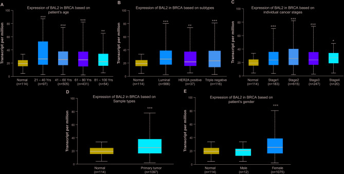
Methods: The expression patterns of BALs were evaluated utilizing multiple databases, including Ualcan, Gene Set Cancer Analysis (GSCA), Search Tool for the Retrieval of Interacting Genes/Proteins (STRING), and Gene Expression Profiling Interactive Analysis (GEPIA). The prognostic significance of BALs was assessed via Kaplan-Meier plotter analysis. Furthermore, the potential mechanisms underlying the contribution of BC progression were predicted through GO and KEGG pathway enrichment analysis. Additionally, the effect of BALs on the malignant behaviors of BC cells was determined using CCK-8 assay, Transwell assay, and TUNEL assay.

Results: The data revealed that the expression levels of both BAL1 and BAL2 were upregulated in BC, whereas no significant change was observed for BAL3. Survival analysis demonstrated a strong association between the overexpression of both BAL1 and BAL2 and favorable prognosis in patients with various subtypes of BC, including estrogen receptor (ER)-positive, ER-negative, Basal, luminal B, HER2-, and HER2 + subtypes. Additionally, the knockdown of BAL1 and BAL2 inhibited the proliferation and migration of BC cells while facilitating apoptosis.

Conclusions: These findings suggest that both BAL1 and BAL2 hold great potential as significant prognostic biomarkers and therapeutic targets for patients with BC.

Keywords: B-aggressive lymphoma protein; Biomarker; Breast cancer; Prognosis.


Conflict of interest statement

Declarations. Ethics approval and consent to participate: This study does not involve animal and human ethics. Consent for publication: Not applicable. Competing interests: The authors declare no competing interests.

Figures

Fig. 1
Fig. 1
Pan-cancer analysis of BAL family expression across 33 cancer types. The expression levels of BAL1 (A), BAL2 (B), and BLA3 (C) in BC tissue samples and normal tissue samples were obtained from the GEPIA database. The red arrows represent BC tissues, while the grey columns represent normal tissues. A total of 1085 BC tissue samples and 291 normal tissue samples were included to investigate BAL expression levels. The y-axis indicates the logarithm of gene expression in the samples. *p < 0.05 indicates a significant difference between normal tissue samples and BC tissue samples. BAL, B-aggressive lymphoma; GEPIA, gene expression profiling interactive analysis
Fig. 2
Fig. 2
Expression profiles of BAL family in BC based on GEPIA database (A) and ENCORI (B) database. The GSCA database comprised 1104 BC tissue samples and 114 normal tissue samples, whereas the ENCORI database included data from 1104 BC tissue samples and 113 normal tissue samples. BAL, B-aggressive lymphoma; BC, breast cancer; GEPIA, gene expression profiling interactive analysis; ENCORI, encyclopedia of RNA interactomes
Fig. 3
Fig. 3
Analysis of BAL1 expression patterns in BC patients with distinct pathologic characteristics. Ualcan analysis revealed the transcriptional levels of BAL1 in BC patients with different age (A), cancer subtypes (B), cancer stages (C), sample types (D) and gender (E). Subtype composition: Normal samples: 114, Luminal subtype: 566, HER2 + subtype: 37, Triple-negative subtype: 116. The statistical significance between each group with normal sample was represented with asterisk (*p < 0.05, **p < 0.01, ***p < 0.001). BAL1, B-aggressive lymphoma 1; BC, breast cancer; Ualcan, University of Alabama cancer
Fig. 4
Fig. 4
Analysis of BAL2 expression patterns in BC patients with distinct pathologic characteristics. Ualcan analysis revealed the transcriptional levels of BAL2 in BC patients with different age (A), cancer subtypes (B), cancer stages (C), sample types (D) and gender (E). Subtype composition: Normal samples: 114, Luminal subtype: 566, HER2 + subtype: 37, Triple-negative subtype: 116. The statistical significance between each group with normal sample was represented with asterisk (*p < 0.05, **p < 0.01, ***p < 0.001). BAL2, B-aggressive lymphoma 2; BC, breast cancer; Ualcan, University of Alabama cancer
Fig. 5
Fig. 5
Prognostic significance of BAL1 expression in BC patients with different molecular subtypes. Kaplan-Meier plots were conducted to draw the RFS curves for all patients (n = 2032) (A), ER-positive (n = 1417) (B), ER-negative (n = 615) (C), Basal-like (n = 442) (D), Luminal A-like (n = 631) (E), Luminal B-like (n = 566) (F), HER2-negative (n = 1571) (G) and HER2-positive (n = 461) (H) subtypes. BAL1, B-aggressive lymphoma 1; BC, breast cancer; ER, estrogen receptor; HER2, human epidermal growth factor receptor 2
Fig. 6
Fig. 6
Prognostic significance of BAL2 expression in BC patients with different molecular subtypes. Kaplan-Meier plots were conducted to draw the RFS curves for all patients (n = 2032) (A), ER-positive (n = 1417) (B), ER-negative (n = 615) (C), Basal-like (n = 442) (D), Luminal A-like (n = 631) (E), Luminal B-like (n = 566) (F), HER2-negative (n = 1571) (G) and HER2-positive (n = 461) (H) subtypes. BAL2, B-aggressive lymphoma 2; BC, breast cancer; ER, estrogen receptor; HER2, human epidermal growth factor receptor 2
Fig. 7
Fig. 7
Protein interaction network and functional annotation analysis of BALs in BC. A Protein-protein interaction network of BALs and their associated co-expressed genes was conducted by STRING analysis. B GO and KEGG pathway enrichment analysis on SRplot platform was performed to predict the potential functions of BALs and their co-expressed proteins, including biological process, cellular component, molecular function, as well as pathway. BAL, B-aggressive lymphoma; BC, breast cancer; STRING, search tool for the retrieval of interacting genes/proteins; GO, gene ontology; KEGG, Kyoto encyclopedia of genes and genomes
Fig. 8
Fig. 8
Effect of BAL1 and BAL2 on malignant behaviors of MCF-7 cells. MCF-7 cells were transfected with siNC, siBAL1-1, siBAL1-2, siBAL2-1, or siBAL2-2 and subsequently subjected to RT-qPCR assay (A), Western blot assay (B), CCK-8 assay (C), Transwell assay (D), and TUNEL assay (E). Each experiment was repeated at least three times. Data are presented as means ± SDs from three independent experiments *p < 0.05. BAL1, B-aggressive lymphoma 1; BAL2, B-aggressive lymphoma 2; BC, breast cancer; RT-qPCR, real-time quantitative polymerase chain reaction; CCK-8, cell counting kit-8
Fig. 9
Fig. 9
Effect of BAL1 and BAL2 on malignant behaviors of MDA-MB-231 cells. MDA-MB-231 cells were transfected with siNC, siBAL1-1, siBAL1-2, siBAL2-1, or siBAL2-2 and subsequently subjected to Western blot assay (A), CCK-8 assay (B), Transwell assay (C), and TUNEL assay (D). Each experiment was repeated at least three times. Data are presented as means ± SDs from three independent experiments **p < 0.01, ***p < 0.001, ****p < 0.0001. BAL1, B-aggressive lymphoma 1; BAL2, B-aggressive lymphoma 2; BC, breast cancer; RT-qPCR, real-time quantitative polymerase chain reaction; CCK-8, cell counting kit-8

Similar articles

References

    1. Bray F, Laversanne M, Sung H, Ferlay J, Siegel RL, Soerjomataram I, et al. Global cancer statistics 2022: GLOBOCAN estimates of incidence and mortality worldwide for 36 cancers in 185 countries. Cancer J Clin. 2024;74(3):229–63. - PubMed
    1. Azamjah N, Soltan-Zadeh Y, Zayeri F. Global trend of breast cancer mortality rate: a 25-year study. Asian Pac J Cancer Prev. 2019;20(7):2015–20. - PMC - PubMed
    1. Lu H, Shi C, Liu X, Liang C, Yang C, Wan X, et al. Identification of ZG16B as a prognostic biomarker in breast cancer. Open Med (Wars). 2021;16(1):1–13. - PMC - PubMed
    1. Wilkinson L, Gathani T. Understanding breast cancer as a global health concern. Br J Radiol. 2022;95(1130):20211033. - PMC - PubMed
    1. Liu Y, Li Y, Du C, Kuang S, Zhou X, Zhang J, et al. Underlying mechanisms of epithelial splicing regulatory proteins in cancer progression. J Mol Med (Berl). 2022;100(11):1539–56. - PubMed

Substances