. 2025 Jul 10:12:1614342.
doi: 10.3389/fmed.2025.1614342. eCollection 2025.

Identification and validation of mitochondria-related genes in panvascular diseases

Affiliations

Identification and validation of mitochondria-related genes in panvascular diseases

Yingfen Li et al. Front Med (Lausanne). .

Abstract

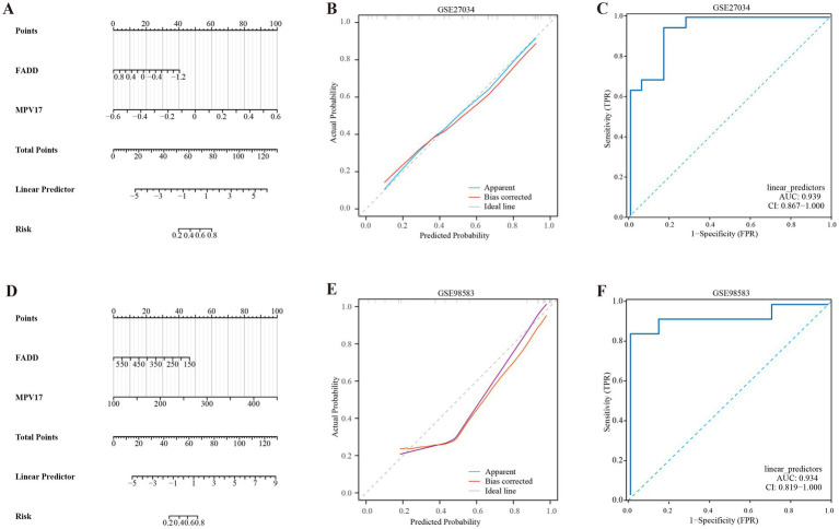
Panvascular diseases represent a spectrum of vascular conditions where atherosclerosis plays a central role in the pathophysiology. This study focused on identifying differentially expressed genes (DEGs) related to mitochondria and key genes associated with peripheral artery disease (PAD) and coronary artery disease (CAD). This study identified MPV17 as a key mitochondrial gene bridging peripheral artery disease (PAD) and coronary artery disease (CAD). Analysis of GEO datasets revealed differentially expressed mitochondrial genes, with MPV17, FADD, HLCS, and PEX3 highlighted. A diagnostic nomogram, developed using LASSO and Random Forest methods, demonstrated high accuracy in predicting PAD and CAD (AUC >0.93). Furthermore, the study revealed significant alterations in immune cell infiltration associated with both diseases, suggesting a potential role for immune modulation in panvascular disease. MPV17 shows promise as a diagnostic marker for early identification and differentiation of these vascular conditions.

Keywords: coronary artery disease; immune infiltration; mitochondria-related genes; panvascular diseases; peripheral artery disease.


Conflict of interest statement

The authors declare that the research was conducted in the absence of any commercial or financial relationships that could be construed as a potential conflict of interest.

Figures

Figure 1
Figure 1
Flowchart of the study design.
Figure 2
Figure 2
Identification of differentially expressed genes (DEGs). DEG heatmaps and volcano plots for the (A,B) PAD and (C,D) CAD datasets.
Figure 3
Figure 3
Identification of mitochondria-related DEGs and functional enrichment between PAD and CAD. (A) Venn diagram of the DEGs. (B) Venn diagram of the mitochondria-related DEGs. (C) GO and KEGG enrichment analyses of DEGs with the same expression trends. (D) GO and KEGG enrichment analyses of the mitochondria-related DEGs.
Figure 4
Figure 4
Screening of mitochondria-related hub genes and evaluation of their diagnostic values. (A) LASSO analysis for screening mitochondria-related hub genes in GSE27034. (B) Identification of mitochondria-related hub genes according to the importance of variables by random forest (RF) analysis of GSE27034. (C) LASSO analysis for screening mitochondria-related hub genes in GSE98583. (D) Identification of mitochondria-related hub genes according to the importance of variables by RF analysis of GSE98583. (E) Receiver operating characteristic (ROC) curves of the four hub genes to assess their diagnostic values in the GSE27034 and GSE98583 datasets.
Figure 5
Figure 5
Expression levels of the four hub genes in the GSE27034 and GSE98583 datasets. (A) Expression level of FADD, MPV17, PEX3 and HLCS in GSE27034. (B) Expression level of FADD, MPV17, PEX3 and HLCS in GSE98583. *p < 0.05, **p < 0.01, and ***p < 0.001.
Figure 6
Figure 6
Development of the diagnostic nomogram model. (A) Nomogram predicting the probability of PAD. (B) Calibration curves of the PAD risk models. (C) ROC curve of the PAD risk model. (D) Nomogram predicting the probability of CAD. (E) Calibration curves of the CAD risk models. (F) ROC curve of the CAD risk model.
Figure 7
Figure 7
Immune cell infiltration analyses in PAD and CAD. (A) Boxplot showing the comparison of 22 kinds of immune cells between PAD and the control group. (B) Boxplot showing the comparison of 22 kinds of immune cells between CAD and the control group. (C) Heatmap representing the associations of the differentially infiltrated immune cells with the four hub genes in PAD for the threshold of p < 0.05, *p < 0.05, **p < 0.01, and ***p < 0.001. (D) Heatmap representing the associations of the differentially infiltrated immune cells with the four hub genes in CAD for the threshold of p < 0.05, *p < 0.05, **p < 0.01, and ***p < 0.001.
Figure 8
Figure 8
Gene set enrichment analysis (GSEA) for MPV17 in (A) PAD and (B) CAD.

Similar articles

References

    1. Chinese College of Cardiovascular Physicians . Chinese expert consensus on antithrombotic therapy of panvascular diseases (2024 edition). Zhonghua Yi Xue Za Zhi. (2024) 104:906–23. doi: 10.3760/cma.j.cn112137-20231101-00959, PMID: - DOI - PubMed
    1. Hu Y, Zhao Y, Li P, Lu H, Li H, Ge J. Hypoxia and panvascular diseases: exploring the role of hypoxia-inducible factors in vascular smooth muscle cells under panvascular pathologies. Sci Bull. (2023) 68:1954–74. doi: 10.1016/j.scib.2023.07.032, PMID: - DOI - PubMed
    1. Zhou X, Yu L, Zhao Y, Ge J. Panvascular medicine: an emerging discipline focusing on atherosclerotic diseases. Eur Heart J. (2022) 43:4528–31. doi: 10.1093/eurheartj/ehac448, PMID: - DOI - PubMed
    1. Chan AW. Expanding roles of the cardiovascular specialists in panvascular disease prevention and treatment. Can J Cardiol. (2004) 20:535–44. - PubMed
    1. Hu Y, Zhao Y, Dai N, Lu H, Ge J. Unwavering excellence: how to be a competent cardiovascular doctor in “panvascular medicine +”. Innovation. (2023) 4:100489. doi: 10.1016/j.xinn.2023.100489, PMID: - DOI - PMC - PubMed

LinkOut - more resources