. 2025 Jun 20:12:rbaf062.
doi: 10.1093/rb/rbaf062. eCollection 2025.

Two-in-one: multifunctional poloxamer hydrogel accelerates endometrial regeneration and fertility restoration through synergistic regulation of KGF-2 and NO

Affiliations

Two-in-one: multifunctional poloxamer hydrogel accelerates endometrial regeneration and fertility restoration through synergistic regulation of KGF-2 and NO

Yijia Zhang et al. Regen Biomater. .

Abstract

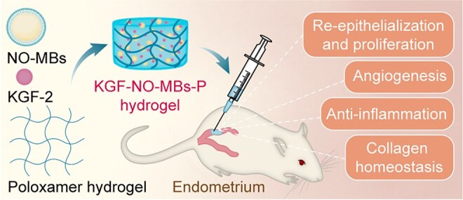
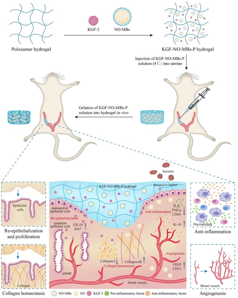
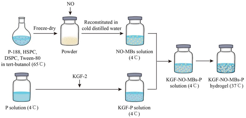
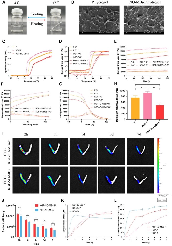
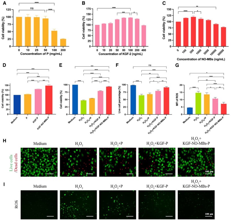
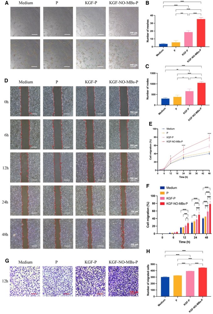
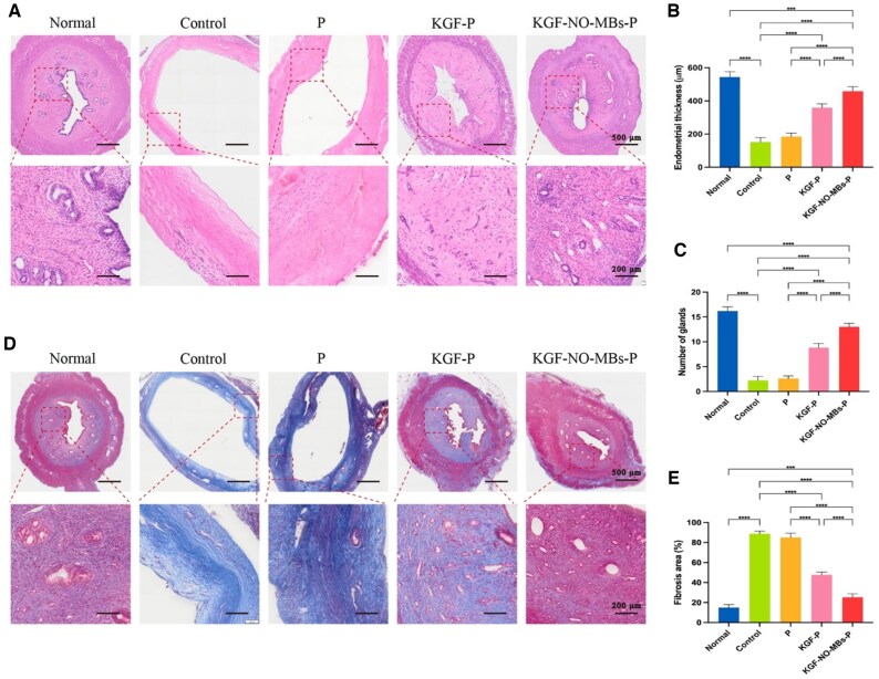
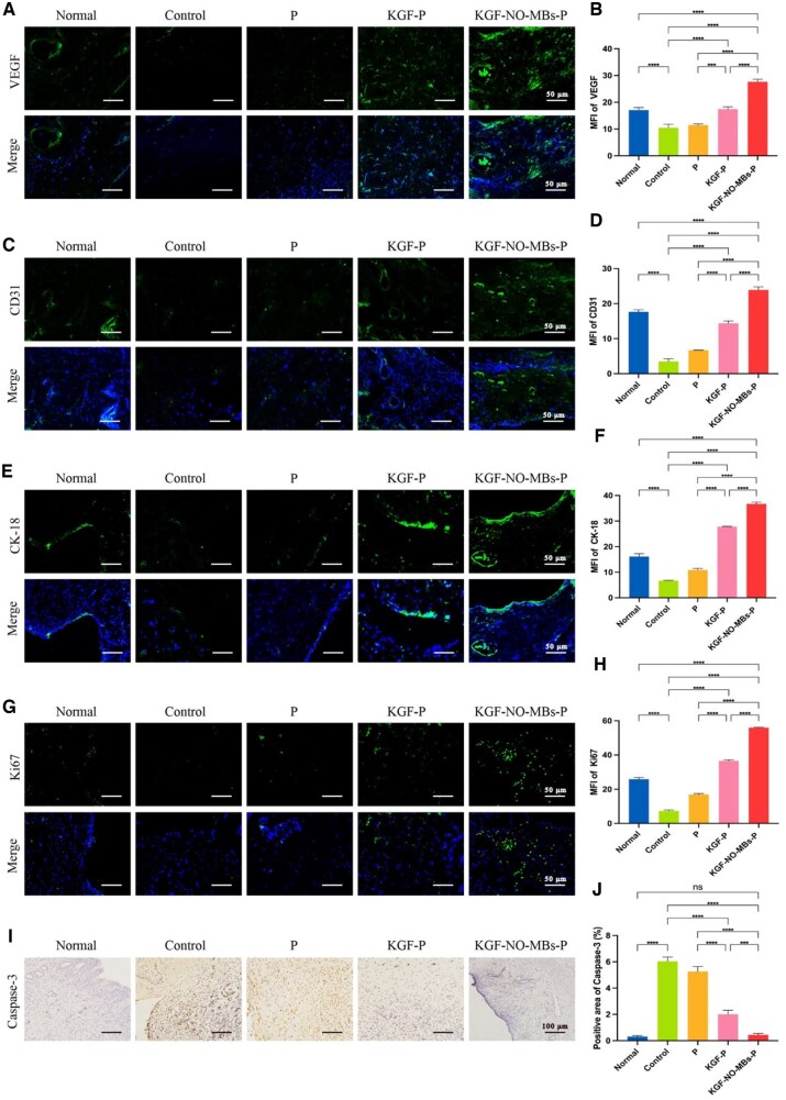
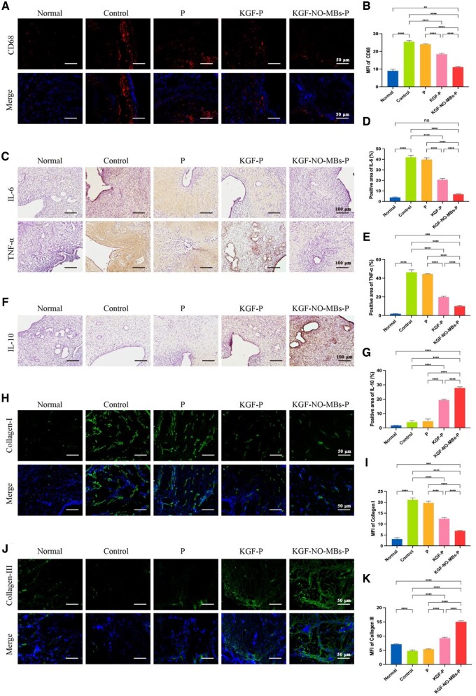
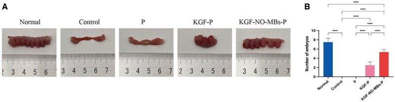
A healthy endometrium is crucial for embryo implantation and pregnancy maintenance. Thin endometrium, reduced glands and fibrosis resulting from infection or mechanical injury, are the primary causes of long-term infertility and poor pregnancy outcomes. Unfortunately, these issues have not been resolved by conventional clinical methods. Keratinocyte growth factor-2 (KGF-2) is an epithelial mitogen that regulates proliferation and migration of epithelial cells. Nitric oxide (NO) is involved in maintaining vascular homeostasis and angiogenesis. Poloxamer-407 (P) hydrogel is a promising topical drug delivery system due to its excellent solution-gel transition properties in response to body temperature. In this study, therapeutic NO gas was first prepared into stabilized microbubbles (NO-MBs). Subsequently, KGF-2 and NO-MBs were encapsulated into micelles of P hydrogel to form a multifunctional temperature-sensitive (28.9-31.8°C) hydrogel (KGF-NO-MBs-P hydrogel). This hydrogel not only exhibited suitable apparent viscosity, bio-adhesive and mechanical properties for application in situ but also showed sustained release of KGF-2 and NO. In vivo, KGF-NO-MBs-P hydrogel effectively restored endometrial morphology, increased the number of glands and endometrial thickness, reversed endometrial fibrosis and improved pregnancy outcomes by synergistic regulation of KGF-2 and NO. Repair of endometrial injury was closely related to promoting neovascularization, inducing endometrial cell proliferation and epithelialization, inhibiting apoptosis and inflammation and balancing collagen subtypes. Therefore, KGF-NO-MBs-P hydrogel may be useful in promoting endometrial regeneration and fertility restoration through in situ microinjection. This study represented a convenient, safe and promising method for repair of endometrial injury.

Keywords: endometrial injury; keratinocyte growth factor; nitric oxide; poloxamer hydrogel; regenerative repair.


Figures

None
Graphical abstract
Figure 1.
Figure 1.
Schematic illustration of applying KGF-NO-MBs-P hydrogel for regeneration of injured endometrium.
Figure 2.
Figure 2.
Preparation of KGF-NO-MBs-P hydrogel.
Figure 3.
Figure 3.
Characterization of NO-MBs and NO-MBs-P hydrogel. (A) Macroscopic appearance of cold NO-MBs solution and NO-MBs-P hydrogel. (B) NO-MBs in NO-MBs solution and NO-MBs-P hydrogel under optical microscopy. (C) Diagram of NO-MBs. (D and E) Microscopy images and numbers of NO-MBs in NO-MBs solution and NO-MBs-P hydrogel. (F) Apparent viscosity–temperature curves of NO-MBs-P hydrogel. (G) Maximum adhesive force of NO-MBs-P hydrogel. (H) NO release curves of NO-MBs-P hydrogel. *P < 0.05, **P < 0.01, ***P < 0.001, ****P < 0.0001, ns, not significant.
Figure 4.
Figure 4.
Characterization of KGF-NO-MBs-P hydrogel. (A) Macroscopic appearance of P hydrogel. (B) SEM images of P hydrogel and NO-MBs-P hydrogel. (C) Apparent viscosity–temperature curves of P hydrogel, KGF-P hydrogel and KGF-NO-MBs-P hydrogel. (D–G) G′ and G″ of temperature sweep, time sweep, frequency sweep and strain sweep of P hydrogel, KGF-P hydrogel and KGF-NO-MBs-P hydrogel. (H) Maximum adhesive force of P hydrogel, KGF-P hydrogel and KGF-NO-MBs-P hydrogel. (I and J) Fluorescence images and intensity of FITC-KGF-NO-MBs solution and FITC-KGF-NO-MBs-P hydrogel in uterus. (K) NO release curves of NO-MBs solution, NO-MBs-P hydrogel and KGF-NO-MBs-P hydrogel. (L) KGF-2 release curves of KGF-2 solution, KGF-P hydrogel and KGF-NO-MBs-P hydrogel. *P < 0.05, **P < 0.01, ***P < 0.001, ****P < 0.0001, ns, not significant.
Figure 5.
Figure 5.
In vitro cytotoxicity and effect of KGF-NO-MBs-P hydrogel. (A–C) Cell viability of HUVECs treated with P-407, KGF-2 and NO-MBs (combined with 100 ng/mL KGF-2). (D) Cell viability of HUVECs treated with P hydrogel, KGF-P hydrogel and KGF-NO-MBs-P hydrogel. (E) Cell viability of HUVECs treated with P hydrogel, KGF-P hydrogel and KGF-NO-MBs-P hydrogel after H2O2 pretreatment. (F and H) Quantitative analysis and fluorescence images of live cells treated with P hydrogel, KGF-P hydrogel and KGF-NO-MBs-P hydrogel after H2O2 pretreatment. (G and I) Fluorescence intensity and images of ROS in HUVECs treated with P hydrogel, KGF-P hydrogel and KGF-NO-MBs-P hydrogel after H2O2 pretreatment. *P < 0.05, **P < 0.01, ***P < 0.001, ****P < 0.0001, ns, not significant.
Figure 6.
Figure 6.
The impact of KGF-NO-MBs-P hydrogel on tube formation and cell migration. (A) Tube formation assay images of HUVECs treated with P hydrogel, KGF-P hydrogel and KGF-NO-MBs-P hydrogel. (B and C) Quantitative analysis of meshes and nodes. (D) Cell scratch assay images of HUVECs treated with P hydrogel, KGF-P hydrogel and KGF-NO-MBs-P hydrogel. (E and F) Analysis of cell migration by measuring scratch areas. (G) Transwell migration assay images of HUVECs treated with P hydrogel, KGF-P hydrogel and KGF-NO-MBs-P hydrogel. (H) Quantitative analysis of migrated cells. *P < 0.05, **P < 0.01, ***P < 0.001, ****P < 0.0001, ns, not significant.
Figure 7.
Figure 7.
Assessment of treatment-related impact in uterine morphology and fertility. (A) H&E staining images of uterus in each group. (B and C) Quantitative analysis of endometrial thickness and number of glands. (D) Masson's staining images of uterus in each group. (E) Relative analysis of fibrosis areas. *P < 0.05, **P < 0.01, ***P < 0.001, ****P < 0.0001, ns, not significant.
Figure 8.
Figure 8.
Administration of KGF-NO-MBs-P hydrogel promoted angiogenesis, epithelium proliferation and inhibited apoptosis. (A and B) Immunofluorescent staining images and mean fluorescence intensity (MFI) of VEGF in each group. (C and D) Immunofluorescent staining images and MFI of CD31 in each group. (E and F) Immunofluorescent staining images and MFI of CK-18 in each group. (G and H) Immunofluorescent staining images and MFI of Ki67 in each group. (I and J) Immunohistochemical staining images and quantitative analysis of Caspase-3 in each group. *P < 0.05, **P < 0.01, ***P < 0.001, ****P < 0.0001, ns, not significant.
Figure 9.
Figure 9.
Administration of KGF-NO-MBs-P hydrogel inhibited inflammation and maintained extracellular matrix homeostasis. (A and B) Immunofluorescent staining images and MFI of CD68 in each group. (C–E) Immunohistochemical staining images and quantitative analysis of IL-6 and TNF-α in each group. (F and G) Immunohistochemical staining images and quantitative analysis of IL-10 in each group. (H and I) Immunofluorescent staining images and MFI of Collagen-I in each group. (J and K) Immunofluorescent staining images and MFI of Collagen-III in each group. *P < 0.05, **P < 0.01, ***P < 0.001, ****P < 0.0001, ns, not significant.
Figure 10.
Figure 10.
Assessment of treatment-related impact in fertility. (A) Images of uterus with embryo implantation in each group. (B) Quantitative analysis of embryo numbers. *P < 0.05, **P < 0.01, ***P < 0.001, ****P < 0.0001, ns, not significant.

Similar articles

References

    1. Zhengli Z, Xiaomei W, Tingwei C, Bo Z, Wenxin L, Min Z, Jingxue Z, Dong E, Tianqing L. Restoration of functional endometrium in an intrauterine adhesion rat model with endometrial stromal cells transplantation. Stem Cell Res Ther 2024;15:181. - PMC - PubMed
    1. Siyu X, Ming W, Xinhao Z, Xiu Z, Lina Y, Kang Z, Yiyi K, Jun L, Yunci Z, Wang W, Dirong D, Hong C, Hui L. Treating intrauterine adhesion using conditionally reprogrammed physiological endometrial epithelial cells. Stem Cell Res Ther 2022;13:178. - PMC - PubMed
    1. Zhao Y, Chen S, Huang Q, Chen W, Xia T, Shi Y, Gao H, Shi Q, Lin S. Repair abilities of mouse autologous adipose-derived stem cells and ShakeGel™3D complex local injection with intrauterine adhesion by BMP7-Smad5 signaling pathway activation. Stem Cell Res Ther 2021;12:191. - PMC - PubMed
    1. Hwang S-Y, Lee D, Lee G, Ahn J, Lee Y-G, Koo HS, Kang Y-J. Endometrial organoids: a reservoir of functional mitochondria for uterine repair. Theranostics 2024;14:954–72. - PMC - PubMed
    1. Zhang H, Zhang Q, Zhang J, Sheng F, Wu S, Yang F, Li W. Urinary bladder matrix scaffolds improve endometrial regeneration in a rat model of intrauterine adhesions. Biomater Sci 2020;8:988–96. - PubMed

LinkOut - more resources