. 2025 Jun 8;10(4):e70033.
doi: 10.1002/btm2.70033. eCollection 2025 Jul.

Multiaxial rotational loading compromises the transition zone of the intervertebral disc: Ex vivo study using next-generation bioreactors

Affiliations

Multiaxial rotational loading compromises the transition zone of the intervertebral disc: Ex vivo study using next-generation bioreactors

Amra Šećerović et al. Bioeng Transl Med. .

Abstract

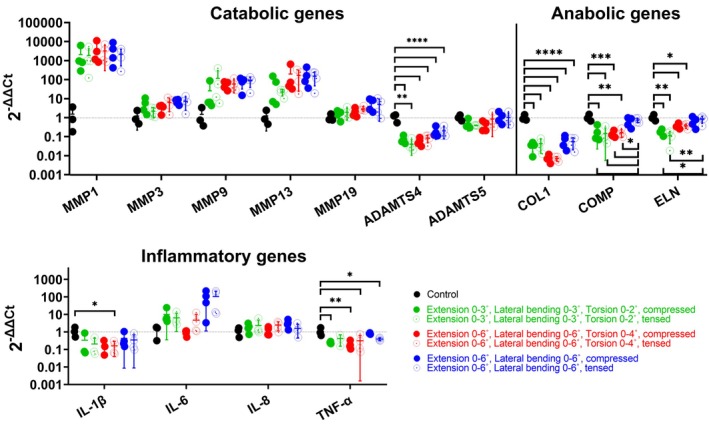
Bioreactors have become indispensable tools in spine research, enabling long-term intervertebral disc culture under controlled biological and mechanical conditions. Conventional systems are often limited to uniaxial loading, restricting their ability to replicate the complex, multidirectional biomechanics of the spine. To overcome this limitation, we developed a next-generation bioreactor capable of simulating multiaxial motions while preserving the disc's biological environment. In this study, we investigated the effects of complex loading patterns on early disc degeneration by subjecting bovine whole-organ discs to combined extension, lateral bending, and torsion at 0.3 Hz for 2 h daily over 14 days. To assess the impact of loading magnitude and the specific contribution of torsion, discs were exposed to either low- or high-angle rotations, with or without torsional loading at higher angles. Histological analysis revealed a marked loss of glycosaminoglycans (GAG) and collagen type II within the inner annulus fibrosus and transitional nucleus pulposus (NP), encompassing the transition zone (TZ), as well as GAG depletion in the central NP. Matrix degradation was observed across all loading conditions, with the most severe breakdown occurring under high-angle extension, bending, and torsion. All loading regimes induced cell death in the TZ and central NP, although torsion-free loading better maintained cell viability. These findings highlight the TZ, alongside the commonly affected NP, as a critical early site of degeneration. The study further underscores the importance of incorporating multiaxial loading in disc degeneration models and provides new insights into the biomechanical mechanisms underlying disc pathology.

Keywords: bioreactors; intervertebral disc; multiaxial loading; transition zone; whole organ culture.


Conflict of interest statement

All authors declare that they have no conflicts of interest with respect to this work.

Figures

FIGURE 1
FIGURE 1
(a) Next‐generation multiaxial bioreactor designed for applying complex mechanical loading and precisely controlling the biological environment of bovine ex vivo whole organ models. The system comprises a hexapod actuator, control software, an incubator, and a previously described chamber with integrated mechanical interfaces and the specimen holder. A close‐up view of the chamber is shown. (b) Illustration of the six degrees of freedom in spine motion that can be simulated using the multiaxial bioreactor. (c) Experimental design and parameters for this study, testing combined rotational spine movements: extension and lateral bending, with or without the addition of torsion. The asymmetrical loading pattern indicates the directly loaded, compressed side of the disc and the counter‐loaded, tensed disc side.
FIGURE 2
FIGURE 2
Disc height changes were measured daily over 14 days, following loading and free‐swelling recovery. Measurements of height after swelling were first recorded on day 2, after the recovery from initial loading on day 1. Changes in height following loading were calculated relative to the pre‐loading height recorded on the same day. Changes in height following swelling were calculated relative to the height before loading recorded the previous day. Data show mean values from 4 samples and standard deviation. A repeated measures two‐way ANOVA test was performed to analyze the effects of loading parameters and time on height changes.
FIGURE 3
FIGURE 3
Analysis of the extracellular matrix in different regions of the intervertebral disc. Panels (a)–(d) show representative macroscopic and close‐up images of a control and a loaded specimen with altered composition from each experimental group, analyzed using different staining methods. Graphs (b)–(d) show the analysis of positive signals from all control and loaded specimens. For loaded groups, data points represent individual measurements obtained from two adjacent locations at the disc center for each sample, except when image quality permitted quantification at only one site. (a) Picrosirius red staining highlights the organized lamellae defining the outer and inner annulus fibrosus (AF), and the randomly organized nucleus pulposus (NP), which includes transitional and central regions. The inner AF and transitional NP encompass the transition zone (TZ). (b) Safranin‐O staining visualizes glycosaminoglycan (GAG) content in red, while fast green counterstains fibrous components. (c), (d) Immunolabeling identifies the distribution of collagen type I and type II. Close‐up images show a loss of GAG and collagen type II signals in the TZ. Arrows indicate vertical tissue cracks within this region, with cells aligned along the cracks (upward arrows) and GAG deposition alongside cells (downward arrows). Scale bars indicate 1 mm. Statistical analysis between all groups was performed using one‐way ANOVA or Kruskal–Wallis test, where p < 0.05 (*) and p < 0.01 (**) were statistically significant.
FIGURE 4
FIGURE 4
Absolute release of glycosaminoglycans (GAG; a) and interleukin‐6 (IL‐6; b) until the first medium change at day 4 (green and red groups) and day 5 (blue group), followed by cumulative release until day 14 of daily loading and free‐swelling periods. Data are presented as mean values from 4 samples and standard deviation. A repeated measures two‐way ANOVA test was performed to analyze the effects of loading parameters and time, where a p < 0.05 (*) was statistically significant between the red and blue groups (a), red and green groups on day 2 (b) and red and green and red and blue groups on day 4 (b).
FIGURE 5
FIGURE 5
Cell viability was visualized and quantified in sections from non‐loaded controls and groups subjected to different loading parameters, stained with lactate dehydrogenase and ethidium homodimer. (a) Representative images show live cells (blue and blue/orange) and dead cells (orange) across a portion of the disc, encompassing the outer and inner annulus fibrosus (AF) and the transitional and central nucleus pulposus (NP). Higher‐magnification images highlight selected regions of interest (scale bar 100 μm). (b) Quantification of cell viability in the regions of interest, including from the directly loaded side in compression and the counter‐loaded side in tension. Data represent the mean ± standard deviation of four loaded samples per group and ten control samples. Central NP data were derived from both counter‐loaded and directly loaded disc regions within the same section. Statistical analysis between loaded and counter‐loaded regions within individual groups was performed using parametric or non‐parametric t‐test. Different groups within the same region were analyzed using one‐way ANOVA or the Kruskal–Wallis test, where p < 0.05 (*), p < 0.01 (**), p < 0.001 (***) and p < 0.0001 (****) were statistically significant.
FIGURE 6
FIGURE 6
Expression of catabolic, anabolic, and inflammatory genes in the outer annulus fibrosus loaded in compression or tension and control samples. Gene expression was quantified using the comparative Ct method (ΔΔCt) normalized to an endogenous control gene and day 0 control samples. Samples below the detection limit were not shown on the graph. Statistical analysis between loaded groups, controls, and loaded groups, and compressed and tensed regions of individual groups was performed using the one‐way ANOVA or Kruskal–Wallis tests, where p < 0.05 (*), p < 0.01 (**), p < 0.001 (***) and p < 0.0001 (****) were statistically significant.

Similar articles

References

    1. Lazaro‐Pacheco D, Mohseni M, Rudd S, Cooper‐White J, Holsgrove TP. The role of biomechanical factors in models of intervertebral disc degeneration across multiple length scales. APL Bioeng. 2023;7(2):021501. doi: 10.1063/5.0137698 - DOI - PMC - PubMed
    1. Stokes IAF, Iatridis JC. Mechanical conditions that accelerate intervertebral disc degeneration: overload versus immobilization. Spine. 2004;29(23):2724‐2732. doi: 10.1097/01.brs.0000146049.52152.da - DOI - PMC - PubMed
    1. Gantenbein B, Illien‐Jünger S, Chan S, et al. Organ culture bioreactors—platforms to study human intervertebral disc degeneration and regenerative therapy. Curr Stem Cell Res Ther. 2015;10(4):339‐352. doi: 10.2174/1574888X10666150312102948 - DOI - PMC - PubMed
    1. McKinley JP, O'Connell GD. Review of state‐of‐the‐art micro and macro‐bioreactors for the intervertebral disc. J Biomech. 2024;165:111964. doi: 10.1016/j.jbiomech.2024.111964 - DOI - PubMed
    1. Pfannkuche J‐J, Guo W, Cui S, et al. Intervertebral disc organ culture for the investigation of disc pathology and regeneration—benefits, limitations, and future directions of bioreactors. Connect Tissue Res. 2020;61(3–4):304‐321. doi: 10.1080/03008207.2019.1665652 - DOI - PubMed

LinkOut - more resources