. 2025 Jul 17:2025:1159325.
doi: 10.1155/jdr/1159325. eCollection 2025.

Costunolide Reduces DN Inflammatory Response and Renal Thrombosis by Inhibiting NET Formation

Affiliations

Costunolide Reduces DN Inflammatory Response and Renal Thrombosis by Inhibiting NET Formation

Xiangjing Wang et al. J Diabetes Res. .

Abstract

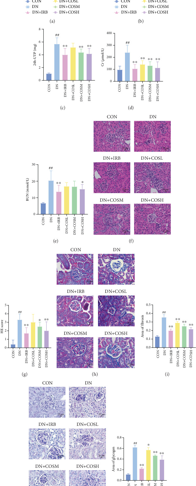
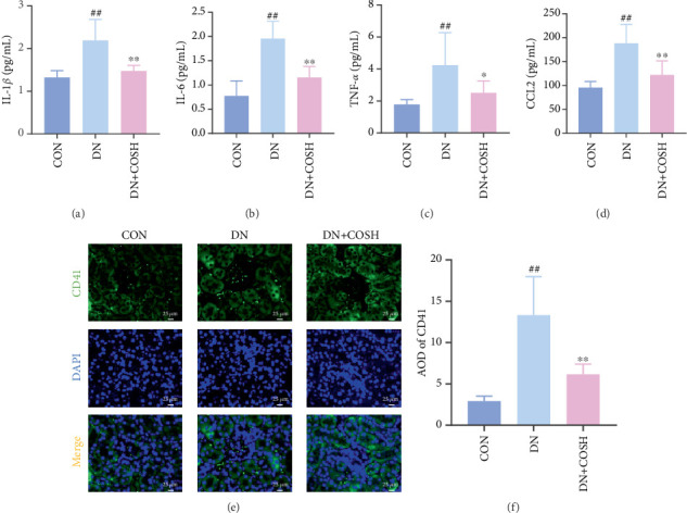
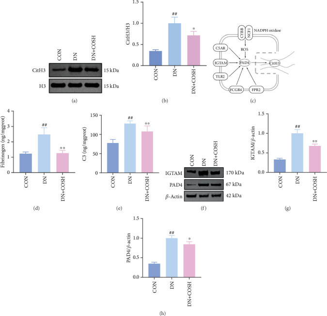
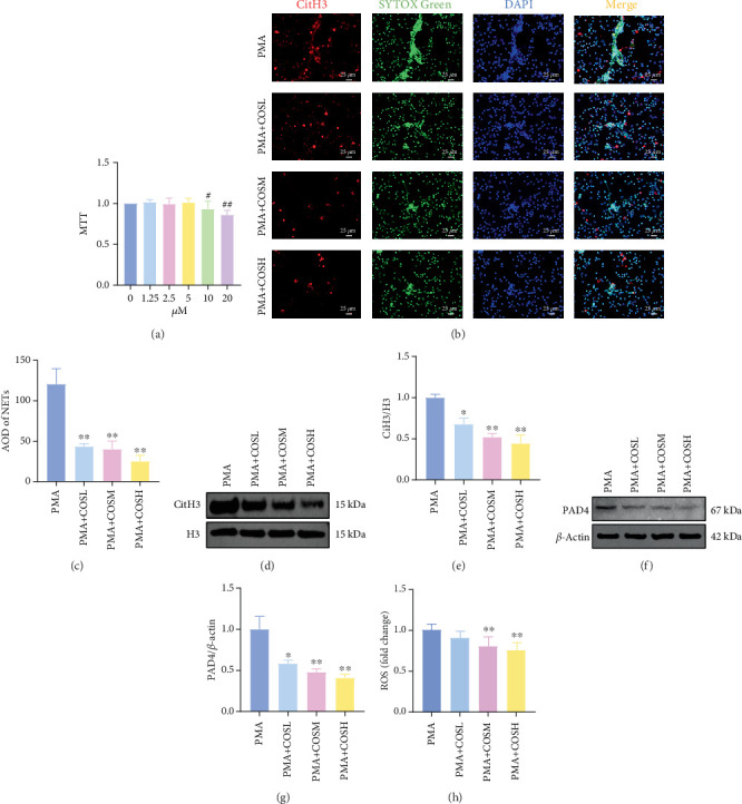
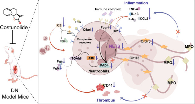
Background: Diabetic nephropathy (DN), a prevalent microvascular complication of diabetes, is characterized by chronic inflammation, oxidative stress, and renal thrombosis. This study is aimed at assessing the therapeutic effects of costunolide (COS) on DN and investigating its mechanism of action in reducing inflammation and platelet activation-mediated thrombosis by inhibiting the formation of neutrophil extracellular traps (NETs). Methods: A DN mouse model was established using a high-sugar, high-fat diet combined with streptozotocin (STZ) administration, followed by treatment with varying doses of COS. The efficacy of COS was assessed through renal function indicators, including 24-h urinary protein levels, serum creatinine, and blood urea nitrogen, alongside renal histopathological analyses using hematoxylin-eosin, Masson's trichrome, and periodic acid-Schiff staining. Transcriptomic analysis was performed to identify gene expression changes in renal tissues after COS treatment. Based on transcriptomic findings, the impact of COS on inflammatory and platelet activation-related markers (IL-1β, IL-6, TNF-α, CCL2, and CD41) was further evaluated. Additionally, the expression of NET formation-related factors (MPO, CitH3, IGTAM, PAD4, C3, and fibrinogen) was analyzed using immunofluorescence, western blot, and ELISA. To validate the in vivo findings, isolated neutrophils were treated with COS in vitro to assess its inhibitory effects on NET formation, including markers such as SYTOX Green, CitH3, ROS, and PAD4. Results: COS treatment significantly improved renal function and mitigated histopathological damage in DN mice. Transcriptomic analysis indicated that COS modulated pathways associated with inflammation and platelet activation, including the complement and coagulation cascades, biosynthesis of cofactors, cytokine-cytokine receptor interactions, NET formation, and NOD-like receptor signaling. COS markedly reduced the expression of inflammatory markers (IL-1β, IL-6, TNF-α, and CCL2) and the platelet activation marker CD41 in renal tissues. Moreover, COS decreased the expression of NET-related proteins, including MPO, CitH3, PAD4, IGTAM, C3, and fibrinogen, while lowering the CitH3/H3 ratio. In vitro, COS significantly inhibited PMA-induced NET formation in neutrophils, as evidenced by reduced SYTOX Green + CitH3+ expression and decreased levels of PAD4 and ROS. Conclusion: COS alleviates inflammation and platelet activation-mediated thrombosis in DN mice, potentially by inhibiting excessive NET formation. These findings highlight the therapeutic potential of COS in managing DN.


Conflict of interest statement

The authors declare no conflicts of interest.

Figures

Figure 1
Figure 1
COS intervention improves renal injury in DN mice. DN mice were given COS intervention at various concentrations (10, 20, and 40 mg/kg). The degree of renal injury in each group was assessed via measurement of blood glucose, body weight, renal function, and pathological staining. (a, b) COS intervention significantly reduces (a) blood glucose and increases (b) body weight in DN mice. (c–e) COS intervention improves renal function and reduces levels of (c) 24-UTP, (d) Cr, and (e) BUN in DN mice. (f–k) COS intervention improves renal histopathological injury in DN mice. (f, g) Results of H&E staining show that COS improves renal histopathological morphology. (h, i) Masson staining shows that COS reduces renal tissue fibrosis. (j, k) PAS staining shows that COS reduces renal tissue glycogen deposition. Data are presented as means ± SD. n = 10 per group. ⁣#p < 0.05, ⁣##p < 0.01 versus control group; ⁣p < 0.05, ⁣∗∗p < 0.01 versus DN group.
Figure 2
Figure 2
Transcriptomic analysis results from screening of differential genes after COS intervention in DN mice. Transcriptomic analysis was performed on CON, DN, and DN + COSH groups. Differential genes (DEGs) were screened for DN versus CON and DN + COSH versus DN using |log2(foldchange)| ≥ 1 and padj ≤ 0.05 as criteria, respectively. (a, b) Volcano plots visualizing DEGs. (c–e) KEGG pathway enrichment analysis revealing intersecting pathways of (c) DN versus CON and (d) DN + COSH versus DN, (e) which were enriched in Fgb, Fga, C3, Itgam, C5ar1, Tlr2, and Fcgr4. n = 3 per group.
Figure 3
Figure 3
Reduction of expression of inflammation- and thrombosis-related factors via COS intervention in renal tissues of DN mice. Renal tissues of mice in CON, DN, and DN + COSH groups were collected. Levels of inflammation-related factors in renal tissues were detected by ELISA. Platelet markers in renal tissues were detected by immunofluorescence. (a–d) COS intervention significantly downregulates levels of (a) IL-1β, (b) IL-6, (c) TNF-α, and (d) CCL2 in renal tissues of DN mice and attenuates inflammatory response in DN mice. In addition, (e, f) COS intervention significantly reduces positive expression of CD41 in glomeruli of DN mice and attenuates coagulation dysfunction phenomenon in DN mice. n = 10 per group.
Figure 4
Figure 4
COS intervention reduces expression of DN NET-related factors in renal tissues of DN mice. Renal tissues from CON, DN, and DN + COSH mice were collected. Western blot was used to detect levels of histone guanosine. ELISA and a biochemical kit were used to detect expression of fibrinogen and C3, genes related to NET formation. Western blot was used to detect expression of IGTAM and PAD4 proteins. (a, b) COS intervention inhibits NET formation in renal tissues of DN mice given a reduction in the expression of CitH3/H3 in nuclei of renal tissue cells. (c) Expression of genes associated with NET formation was also reduced, including (d) fibrinogen and (e) C3, as well as reduced expression of proteins including (f, g) IGTAM and (f, h) PAD4. (a, b, f, g, h) n = 3; (d, e) n = 10.
Figure 5
Figure 5
In vitro experiments showing the inhibition of NET formation by COS intervention. Mice peripheral blood neutrophils were extracted. MTT was then used to screen COS intervention dosages on (a) neutrophils. Subsequently, 100 nM PMA was used for 3 h. For in vitro validation, SYTOX Green + CitH3+ expression was detected by immunofluorescence, ROS level was detected by a biochemical kit, and CitH3/H3 in the nucleus and PAD4 protein expression in total protein were detected by western blot. (b–e) COS intervention reduces NET formation after PMA induction, (b, c) decreases SYTOX Green and CitH3 expression, and (d, e) decreases CitH3/H3 protein expression in nucleus proteins. COS intervention also reduces (h) ROS levels and (f, g) PAD4 protein expression in cells. (a) n = 6; (b–g) n = 3; (h) n = 6.
Figure 6
Figure 6
COS alleviates inflammation and renal thrombosis in DN mice, potentially by inhibiting excessive NET formation.

Similar articles

References

    1. Mauricio D., Gratacòs M., Franch-Nadal J. Diabetic Microvascular Disease in Non-Classical Beds: The Hidden Impact Beyond the Retina, the Kidney, and the Peripheral Nerves. Cardiovascular Diabetology . 2023;22(1):p. 314. doi: 10.1186/s12933-023-02056-3. - DOI - PMC - PubMed
    1. Wang H., Liu D., Zheng B., et al. Emerging Role of Ferroptosis in Diabetic Kidney Disease: Molecular Mechanisms and Therapeutic Opportunities. International Journal of Biological Sciences . 2023;19(9):2678–2694. doi: 10.7150/ijbs.81892. - DOI - PMC - PubMed
    1. Jin Q., Liu T., Qiao Y., et al. Oxidative Stress and Inflammation in Diabetic Nephropathy: Role of Polyphenols. Frontiers in Immunology . 2023;14 doi: 10.3389/fimmu.2023.1185317.1185317 - DOI - PMC - PubMed
    1. Alsharidah A. S. Diabetes Mellitus and Diabetic Nephropathy: A Review of the Literature on Hemostatic Changes in Coagulation and Thrombosis. Blood Research . 2022;57(2):101–105. doi: 10.5045/br.2022.2021204. - DOI - PMC - PubMed
    1. Zheng F., Ma L., Li X., et al. Neutrophil Extracellular Traps Induce Glomerular Endothelial Cell Dysfunction and Pyroptosis in Diabetic Kidney Disease. Diabetes . 2022;71(12):2739–2750. doi: 10.2337/db22-0153. - DOI - PubMed

MeSH terms