. 2025 Jul 10:16:1608074.
doi: 10.3389/fimmu.2025.1608074. eCollection 2025.

Single-cycle, pseudotyped reporter influenza virus to facilitate evaluation of treatment strategies for avian influenza, Ebola and other highly infectious diseases in vivo

Affiliations

Single-cycle, pseudotyped reporter influenza virus to facilitate evaluation of treatment strategies for avian influenza, Ebola and other highly infectious diseases in vivo

Tiong Kit Tan et al. Front Immunol. .

Abstract

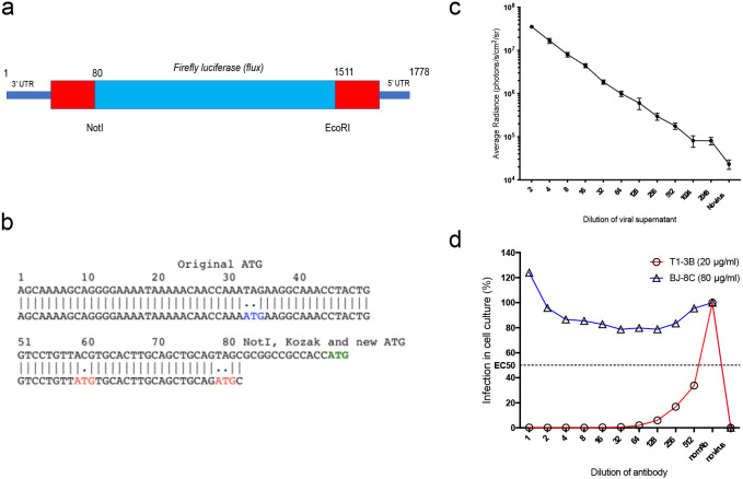
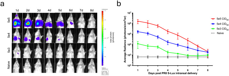
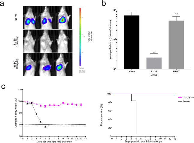
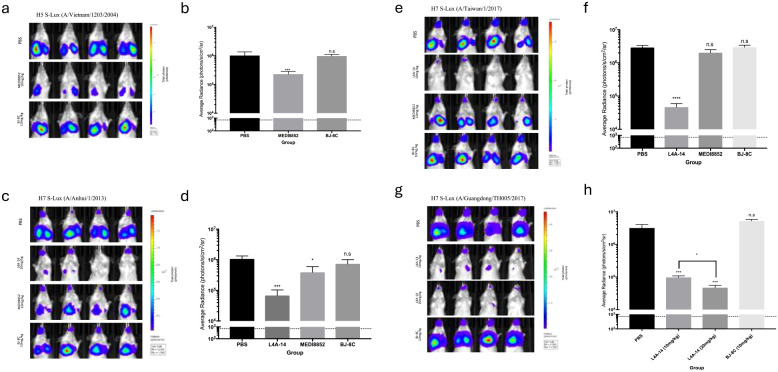
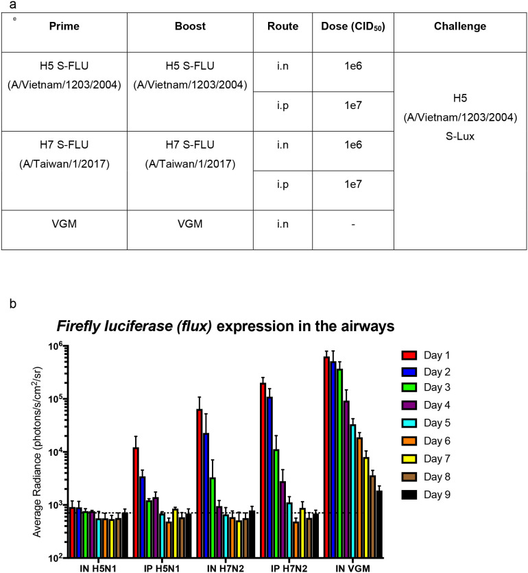
The rapid spread of infectious diseases presents a significant global threat, with seasonal influenza viruses, leading to 290,000-650,000 deaths annually. Emerging high pathogenic influenza strains from animals such as H5N1 and H7N9 further exacerbates pandemic risks. While developing effective vaccines and therapeutics is critical, the evaluation of these interventions is constrained by the requirement for high biosafety containment facilities. To circumvent these challenges, we developed S-Lux, a replication-deficient, single-cycle recombinant influenza virus expressing firefly luciferase (Flux) as a reporter protein. S-Lux can be pseudotyped with haemagglutinin from avian influenza, H5 and H7, enabling real-time monitoring of viral infection in vivo, and facilitate therapeutic antibody evaluation in low-containment facilities. In mice, S-Lux infection resulted in dose-dependent bioluminescent expression in the mouse airways and allowed evaluation of neutralising monoclonal antibodies and clearance of infected cells in mice. To extend this system, we generated ES-Lux by pseudotyping with the Ebola Glycoprotein (GP) and demonstrated that ES-Lux can be used to evaluate the efficacy of Ebola GP-targeting antibodies in vivo. Together, S-Lux and ES-Lux enable robust, simple and time-efficient assessment of antiviral therapy targeting influenza and Ebola virus in vivo, overcoming biosafety constraints that limit traditional efficacy studies.

Keywords: bioluminescence; in vivo imaging; influenza; pandemic; reporter virus.


Conflict of interest statement

The authors declare that the research was conducted in the absence of any commercial or financial relationships that could be construed as a potential conflict of interest.

Figures

Figure 1
Figure 1
Generation and characterisation of S-Lux in vitro. (a) Schematic showing the heamagglutinin virus RNA including 5’ and 3’ Untranslated Regions (UTR) flanking the codon-optimised firefly luciferase (flux) transgene and the position of NotI and EcoRI restriction sites. (b) The construct was based on S-FLU (15) with modifications to the original 3’ packaging sequence, inactivating two ATG codons (depicted in red) and shortening the 5’ packaging sequence from 1275–1778 to 1511–1778 to allow insertion of a larger transgene. A NotI site followed by Kozak sequence were inserted at position 80. The ATG codon (depicted in green) represents the start codon of the inserted flux transgene. (c) Monolayers of MDCK cells seeded in 96-well plates were infected with dilutions of PR8 S-Lux and bioluminescence was measured 24 h post infection. Each data point represents the average of four readings. (d) PR8 S-Lux was incubated with dilutions of a known neutralising antibody (T1-3B), or non-neutralising antibody (BJ-8C), and then added to MDCK cells cultured in 96-well plates. Bioluminescence was measured 24 h post infection. Each data point represents the average of two readings. UTR: untranslated region.
Figure 2
Figure 2
Bioluminescence imaging of mice infected with PR8 S-Lux. The effect of dose response and flux kinetics was imaged in mice (n=4) following i.n. infection with 5e3, 5e4 or 5e5 PR8 S-Lux. (a) Mice were imaged at the indicated time points; images of only one mouse per group per day is shown. (b) Values shown correspond to the average photon flux (photons/s/cm2/sr2) for each treatment group at indicated time points in (a); each data point represents mean ± s.e.m (n=4).
Figure 3
Figure 3
Validation of PR8 S-Lux in vivo using known neutralising antibodies. Mice (n=3) were administered i.p. 10 mg/kg of a known neutralising antibody (T1-3B), a non-relevant antibody (BJ-8C), or PBS at 24 h prior to infection. Mice were then dosed i.n. with ~2e4 CID50 of PR8 S-Lux and (a) imaged at 24 h post infection. (b) Values represent the average photon flux (photons/s/cm2/sr2) as shown in (a); each data point represents mean ± s.e.m (n=3). The statistical significance of differences was calculated using Students’ t-test. (c) Mice (n=6) were treated i.p. with 10 mg/kg of T1-3B at 24 h prior to infection or remained naive. Mice were then dosed i.n. with 1e4 TCID50 (1,000 LD50) of wild type PR8 virus and monitored for weight loss and survival over indicated time point. The statistical significance of differences was calculated using Log-rank test. ***p<0.01, ns, not significant.
Figure 4
Figure 4
Validation of H5 and H7 S-Lux in vivo using known neutralising antibodies. Mice (n=4) were administered i.p. various antibodies or PBS, 24 h prior to i.n. infection with S-Lux pseudotyped with various H5 or H7 strains as indicated, followed by bioluminescence imaging of flux activity 24 h later. Data are presented as average radiance (photons/s/cm2/sr); each data point represents mean ± s.e.m (n=4). (a, b) show mice administered with 10 mg/kg of MEDI8852, non-relevant antibody BJ-8C, or PBS at 24 h prior to infection with 1e6 CID50 of H5 S-Lux (A/Vietnam/1203/2004), and imaged 24 h post-infection. (c, d) show mice administered with 10 mg/kg of MEDI8852, known neutralising antibody L4A-14, non-relevant antibody (BJ-8C), or PBS 24 h prior to infection with 1e6 CID50 of H7 S-Lux (A/Anhui/1/2013), and imaged 24 h post-infection. (e, f) show mice administered with 10 mg/kg of known neutralising antibody L4A-14, non-relevant antibody BJ-8C or PBS at 24 h prior to infection with 1e6 CID50 of H7 S-Lux (A/Taiwan/1/2017), and imaged 24 h post-infection. (g, h) show mice administered with 10 mg/kg or 20 mg/kg of known neutralising antibody (L4A-14), a non-relevant antibody BJ-8C or PBS 24 h prior to infection with 5e5 CID50 of H7 S-Lux (A/Guangdong/TH005/2017), and imaged 24 h post-infection. *p<0.05, ***p<0.001, ****p<0.0001. ns, not significant.
Figure 5
Figure 5
Validation of S-Lux to study lung clearance in vivo post S-FLU immunisation. (a) Table summarising the dosing schedule for the experiment. Mice (n=4-7) were immunised with two doses, two weeks apart, of H5 (A/Vietnam/1203/2004) S-FLU (matched) or H7 (A/Taiwan/1/2017) S-FLU, via either the intranasal (i.n) or intraperitoneal (i.p) route. A control group was included in which the mice were administered i.n with VGM (viral growth media). All mice were then challenged i.n with H5 (A/Vietnam/1203/2004) S-Lux 3 weeks post-immunisation. (b) Mice were imaged daily for up to 9 days post S-Lux infection with data presented as average radiance (photons/s/cm2/sr), mean ± s.e.m (n=4-7).
Figure 6
Figure 6
Validation of Ebola S-Lux (ES-Lux) in vivo using known neutralising antibody. Mice (n=3) were administered i.p with 15 mg/kg of known neutralising antibody KZ52 or PBS 24 h prior to i.v infection with 3.6e5 CID50 of ES-Lux and (a) imaged 24 h post-infection, with (b) data presented as average radiance (photons/s/cm2/sr), mean ± s.e.m (n=3). ****p<0.0001.

References

    1. World Health Organisation . Influenza (Seasonal) Fact Sheets. WHO; (2023). Available online at: https://www.who.int/news-room/fact-sheets/detail/influenza-(seasonal).
    1. Cowling BJ, Jin L, Lau EH, Liao Q, Wu P, Jiang H, et al. Comparative epidemiology of human infections with avian influenza A H7N9 and H5N1 viruses in China: a population-based study of laboratory-confirmed cases. Lancet. (2013) 382:129–37. doi: 10.1016/S0140-6736(13)61171-X, PMID: - DOI - PMC - PubMed
    1. Shi J, Zeng X, Cui P, Yan C, Chen H. Alarming situation of emerging H5 and H7 avian influenza and effective control strategies. Emerg Microbes Infect. (2023) 12:2155072. doi: 10.1080/22221751.2022.2155072, PMID: - DOI - PMC - PubMed
    1. Wang X, Jiang H, Wu P, Uyeki TM, Feng L, Lai S, et al. Epidemiology of avian influenza A H7N9 virus in human beings across five epidemics in mainland China 2013-17: an epidemiological study of laboratory-confirmed case series. Lancet Infect Dis. (2017) 17:822–32. doi: 10.1016/S1473-3099(17)30323-7, PMID: - DOI - PMC - PubMed
    1. Centers For Disease Control and Prevention . Viruses of Special Concern (2023). Available online at: https://www.cdc.gov/flu/pandemic-resources/monitoring/viruses-concern.html (Accessed August 31, 2023).

Substances

LinkOut - more resources