. 2025 Jun 25:15:23.
doi: 10.25259/JCIS_155_2024. eCollection 2025.

Cardiac magnetic resonance imaging in Fabry disease

Affiliations

Cardiac magnetic resonance imaging in Fabry disease

Hatty Hoi Ting Chau et al. J Clin Imaging Sci. .

Abstract

Fabry disease (FD) is a rare X-linked lysosomal storage disorder. Cardiac involvement is frequent in the classic phenotype and late-onset cardiac variant of FD. It is challenging to distinguish FD cardiomyopathy from other forms of unexplained left ventricular hypertrophy, especially in those patients without extracardiac manifestations. Cardiac magnetic resonance imaging is an essential imaging modality for the quantitative and qualitative assessment of FD cardiomyopathy. It helps to monitor disease progress and allows early disease detection in the mild form or subclinical cardiac phenotypes. This review illustrates the characteristic imaging features of FD cardiomyopathy in cardiac MRI, aiming to enhance the awareness of this disease entity among the scope of unexplained cardiomyopathy and promote timely enzyme replacement therapy for patients.

Keywords: Cardiac magnetic resonance imaging; Fabry disease; Hypertrophic cardiomyopathy.


Conflict of interest statement

There are no conflicts of interest.

Figures
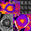
Figure 1:
Figure 1:
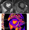
A 46-year-old male patient with known Fabry disease diagnosed 6 years ago. (a) The initial MRI shows diffusely decreased T1 value (883ms*) of left ventricle myocardium with no evidence of left ventricular hypertrophy nor late gadolinium enhancement. (b) Interval MRI shows asymmetrical left ventricular hypertrophy relatively spared the mid inferolateral wall (white arrow), pattern typical for Fabry disease. (c) Latest MRI shows interval development of mild diffuse patchy mid wall enhancement of left ventricle (arrow heads). (d) T1 value of left ventricular mid chamber shows with pseudonormalization of T1 value (1020 ms) without late gadolinium enhancement. (*Machine specific normal range of native T1 value: 1000 ms to 1050 ms.)
Figure 2:
Figure 2:
A 68-year-old female Fabry disease patient with left ventricular hypertrophy . (a) Short-axis cine of left ventricle mid chamber at end diastolic phase shows asymmetrical left ventricular hypertrophy relatively spared the mid chamber inferolateral wall (white arrowheads). (b) Delayed post gadolinium phase-sensitive inversion recovery T1 -weighted sequence shows patchy mid left ventricle most extensive at mid chamber inferolateral wall (white arrow). (c) Native T1 mapping at left ventricular mid chamber short-axis shows pseudonormalization of T1 values at inferolateral wall (1025ms) and decreased T1 values in rest of the left ventricular wall.
Figure 3:
Figure 3:
A 64-year-old male Fabry disease patient with left ventricular hypertrophy. (a) The three chamber view cine at systolic phase shows hypertrophic left ventricular wall, left ventricular outflow tract obstruction with low signal jet at the outflow tract (white arrow) and systolic anterior motion of the mitral valve (white arrowhead). (b) The four chamber view cine at systole (white arrow) demonstrates mitral regurgitation during systole with a low signal jet above the mitral valve.
Figure 4:
Figure 4:
A 67-year-old male Fabry disease patient. Short axis mid chamber view at end diastole shows right ventricular hypertrophy predominantly at inferior and free lateral walls (white arrowheads), while ejection fraction is preserved.
Figure 5:
Figure 5:
A 62-year-old male Fabry disease patient with left ventricular hypertrophy and moderate aortic regurgitation. (a) The three chamber view cine at diastolic phase shows hypertrophic left ventricular wall. A low signal jet seen below the aortic valve (white arrow) during diastole represents regurgitation. (b) The coronal left ventricular outflow tract view shows ectasia of ascending thoracic aorta (measured 3.9cm in calibre on black double arrows) secondary to aortic valve disease.
Figure 6:
Figure 6:
A 67-year-old male Fabry disease patient. (a, b and c) Delayed post gadolinium phase-sensitive inversion recovery T1-weighted sequences in three-chamber, four-chamber and short-axis views show patchy mid wall late gadolinium enhancement in the left ventricular wall predominantly involving the basal inferior and lateral walls (white arrows), which is characteristic of Fabry disease cardiomyopathy.
Figure 7:
Figure 7:
A 50-year-old male Fabry disease patient at storage phase of cardiomyopathy. Native T1 mapping at the mid left ventricular short-axis shows low T1 value (926ms). There is no left ventricular hypertrophy or late gadolinium enhancement in this patient.
Figure 8:
Figure 8:
A 72-year-old male Fabry disease patient in later stage of cardiomyopathy. (a) Delayed post gadolinium phase-sensitive inversion recovery T1 -weighted sequence in mid chamber short-axis view show mid wall late gadolinium enhancement affecting mid inferolateral wall of left ventricle which is not thickened (white arrow). (b and c) Focally elevated T1 value (1118ms) and T2 value (60ms) at the mid chamber inferolateral wall with corresponding mid wall late gadolinium enhancement is suggestive of chronic inflammation. The remaining LV myocardium show diffusely decrease T1 value with normal T2 value.

Similar articles

References

    1. Linhart A, Arad M, Elliott PM, Caforio ALP, Pantazis A, Adler Y. ESC Working Group Myocardial and Pericardial Diseases. Diagnosis and management of cardiac manifestations in Anderson Fabry disease and glycogen storage diseases. Available from: https://www.escardio.org/static-file/escardio/subspecialty/working%20gro... [Last accessed on 2025 Apr 28]
    1. Germain DP. Fabry disease. Orphanet J Rare Dis. 2010;5:30. doi: 10.1186/1750-1172-5-30. - DOI - PMC - PubMed
    1. Umer M, Kalra DK. Cardiac MRI in Fabry disease. Front Cardiovasc Med. 2023;9:1075639. doi: 10.3389/fcvm.2022.1075639. - DOI - PMC - PubMed
    1. MacDermot KD, Holmes A, Miners AH. Anderson-Fabry disease: Clinical manifestations and impact of disease in a cohort of 60 obligate carrier females. J Med Genet. 2001;38:769–75. doi: 10.1136/jmg.38.11.769. - DOI - PMC - PubMed
    1. Niemann M, Herrmann S, Hu K, Breunig F, Strotmann J, Beer M, et al. Differences in Fabry cardiomyopathy between female and male patients: Consequences for diagnostic assessment. JACC Cardiovasc Imaging. 2011;4:592–601. doi: 10.1016/j.jcmg.2011.01.020. - DOI - PubMed

LinkOut - more resources