. 2025 Sep 9;152(10):699-716.
doi: 10.1161/CIRCULATIONAHA.125.073937. Epub 2025 Jul 25.

Junctophilin-2 Regulates Store-Operated Calcium Entry to Drive Cardiac Fibroblast Activation, Fibrotic Repair, and Angiogenesis After Myocardial Infarction

Affiliations

Junctophilin-2 Regulates Store-Operated Calcium Entry to Drive Cardiac Fibroblast Activation, Fibrotic Repair, and Angiogenesis After Myocardial Infarction

Jinxi Wang et al. Circulation. .

Erratum in

Abstract

Background: Calcium (Ca2+) homeostasis in cardiac fibroblasts (CFs) plays a critical role in myocardial repair and remodeling after injury. JPH2 (junctophilin-2; human JPH2 or mouse Jph2) is a structural protein known to regulate intracellular Ca2+ signaling and excitation-contraction coupling in cardiomyocytes. However, the role of JPH2 in CF biology remains unexplored.

Methods: Junctophilin expression was assayed in human and mouse CFs using reverse transcription quantitative polymerase chain reaction, Western blotting, and immunofluorescence. To investigate the functional role of Jph2 in CFs, we assessed Ca2+ handling with live-cell confocal imaging, conducted RNA sequencing analysis, and assayed TGFβ (transforming growth factor β) responses after adenovirus-mediated gene silencing of Jph2 and fibroblast-specific Jph2 knockout. Jph2 interactions in CFs were identified using immunoprecipitation and proximity biotin ligation assays and confirmed by coimmunoprecipitation and rescue experiments. The in vivo role of Jph2 in CFs was investigated in fibroblast-specific Jph2 knockout mice (Col1a2CreERT/Jph2flox/flox, Jph2fKO) at baseline and after myocardial infarction and evaluated for changes in cardiac function, fibrosis, angiogenesis, CF activation, differentiation, and proliferation.

Results: Jph2 was identified as the only junctophilin expressed in CFs and acutely upregulated in response to myocardial infarction. Cellular and RNA sequencing analyses revealed that isolated Jph2-deficient CFs exhibited impaired fibroblast activation, reduced extracellular matrix production, and diminished expression of VEGFA (vascular endothelial growth factor A), VEGFB, and VEGFC after TGFβ treatment. In vivo, Jph2fKO mice displayed exacerbated adverse cardiac remodeling after myocardial infarction, characterized by decreased CF activation/extracellular matrix production, enhanced CF proliferation, worsened systolic dysfunction, increased left ventricular dilation, impaired scar maturation, and decreased angiogenesis. Mechanistically, biochemical assays demonstrated that Jph2 interacts directly with the coiled-coil 2 domain of Stim1 (stromal interaction molecule 1) via its joining region. Jph2 knockdown led to Stim1 protein destabilization, defective store-operated Ca2+entry, and a reduction in both canonical and noncanonical TGFβ signaling pathways. Stim1 overexpression partially rescued the sensitivity of Jph2-deficient CFs to TGFβ, as evidenced by increased expression of periostin and fibronectin-1, as well as enhanced Smad3 phosphorylation.

Conclusions: Our results demonstrate that Jph2 is required in CFs to orchestrate Ca2+ homeostasis, CF activation, and extracellular matrix production and promotes angiogenesis in the infarcted heart, positioning it as a central regulator of cardiac repair after injury.

Keywords: angiogenesis; extracellular matrix; fibroblasts; fibrosis; junctophilin-2; myocardial infarction; store-operated calcium entry.


Conflict of interest statement

Dr Song is an inventor on a patent regarding the use of junctophilin-2 fragments for the treatment of heart failure and other diseases (WO2017214296A1).

Figures

Figure 1.
Figure 1.. Jph2 is the only Junctophilin isoform expressed in mouse and human CFs.
A. RT-PCR data of Jph expression in mouse brain, heart, skeletal muscle and isolated mouse cardiac fibroblasts. B-C. RT-qPCR data of Jph expression from mouse (B, n=5) and human (C, n=6) CFs; D-E. Comparison of Jph2 protein expression in isolated mouse cardiomyocytes (CMs) (n=3) vs. mouse CFs (n=3). F. Immunofluorescence images of Jph2 in a Vimentin-positive CF (bottom) vs. Jph2 in a mouse cardiomyocyte (CM, top panels). Vimentin, a fibroblast marker. Data are presented as mean ± SEM (D). Statistical analyses were conducted using an unpaired t test (D).
Figure 2.
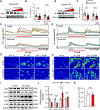
Figure 2.. Jph2 is necessary for Stim1 integrity and SOCE function in CFs.
A. Western blot analysis of Jph2 knockdown efficiency in mouse CFs with increasing amounts of Ad-shJph2 adenovirus vs. negative control virus (Ad-NC). B-D. Quantitation of intracellular Ca2+ amplitudes (B) with exemplar intracellular Ca2+ traces (C) and confocal images (D) measuring SOCE in transduced mouse CFs. Intracellular Ca2+ stores were depleted with thapsigargin (TG) (1 μM) in Ca2+-free solution, followed by the induction of SOCE by perfusing cells with 1.8 mM Ca2+-containing solution. Bar graph in (B) shows averaged peak Ca2+ amplitude of 40–41 cells from 3 independent mouse CF isolations. Traces in (C) show the Ca2+ transients from 15 cells during the treatment protocol. E-H. Similar to panel A-D, but assayed in human CFs transduced with Ad-shJph2 and Ad-NC. Bar graph in (F) shows averaged peak Ca2+ amplitude of 41–52 human CFs from 3 independent experimental repeats. Traces in (G) show the Ca2+ transients from 15 cells. I-J. Western blots (I) and quantitation (J) of mouse CFs showing Jph2 knockdown decreases Stim1 stability and causes a compensatory increase in Orai1 expression (n=3). K. Stim1 gene expression in mouse cardiac fibroblasts infected with Ad-NC and Ad-shJph2, as determined by RNA-sequencing analysis (n=3). Data are presented as mean ± SEM (B, F, J and K). Statistical analyses were conducted using an unpaired t test (B, F, J and K).
Figure 3.
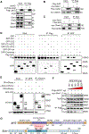
Figure 3.. Jph2 interacts with Stim1 to preserve its stability.
A. Immunoprecipitation assay for Jph2 interacting proteins in CF lysates from WT mice. Cells were infected with negative control (Ad-NC) or Flag-tagged Jph2 (Ad-Flag-Jph2) expressing adenoviruses for 48 hours. B. Co-immunoprecipitation assay using Flag antibody in myc-tagged Stim1 overexpressed HEK293 cells with or without Flag-tagged Jph2-overexpression. C, Co-immunoprecipitation assay using anti-myc antibody in Flag-tagged Jph2 overexpressed HEK293 cells with or without myc-tagged STIM1-overexpression. D. GFP co-IPs from HEK cells co-transfected with GFP-Stim1 cytoplasmic fragments and full-length Flag-tagged Jph2. E. mCherry co-IPs from HEK cells co-transfected with mCherry-Jph2 cytoplasmic flexible fragments and the GFP-Stim1 CC2 domain. CC, coiled-coil; JR, joining region; DR, divergent region; MORN, membrane occupation and recognition nexus motif; P, pseudo-MORN. F. HEK293T cells were transfected with 1000 ng of Flag-Jph2, 1000 ng myc-Stim1, and 100 ng to 2000 ng of GFP-CC2 (fragment of Stim1) plasmids as indicated. Lysates (input; top) were processed with Flag-tagged magnetic beads to pull down Jph2 followed by Western-blotting to determine the relative ability of GFP-CC2 to compete with full length myc-Stim1 for binding Jph2 (bottom). G. Schematic of Jph2 and Stim1 domain structure and interaction domains.
Figure 4.
Figure 4.. RNA sequencing reveals Jph2 is critical for processes involved in CF activation.
A. Experimental design in which primary mouse CFs were infected with adenoviruses for 72 hours prior to RNA isolation and RNA-seq analysis. B and C. RNA-seq volcano plot of differentially expression genes (DEGs) after Ad-shJph2 (B) or Ad-Jph2 (C) infection relative to Ad-GFP infected CFs. Significant DEGs meeting thresholds of >1.5 fold change (0.585 absolute log2 fold change), Benjamini-Hochberg pAdj <0.05, and >1 transcript per million are indicated by darker symbols. D. Venn diagram of significant up- and down-regulated DEGs of Ad-shJph2 and Ad-Jph2 samples relative to Ad-GFP. E and F. Bubble plot of top ten enriched Canonical Pathway terms of each pairwise comparison for significant DEGs using Ingenuity Pathway Analysis. Z-scores <−2 and >+2 delineate thresholds for inactivated and activated terms, respectively. G. Heatmap of k-means clustering of all significant DEGs. H. Heatmap of DEGs commonly associated with fibrosis, TGFβ signaling, and cell cycle progression.
Figure 5.
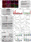
Figure 5.. Jph2 is upregulated in myofibroblasts and is essential for CF activation and differentiation.
A. Immunofluorescence of Jph2 in mouse hearts at baseline (sham) and different days (1, 3, 7) after MI surgery. Top panel: Jph2 (red) and DAPI (blue); Bottom panel: Jph2 (red), Vimentin (green) and DAPI (blue). B-C. TGFβ treatment in mouse CFs induces Jph2 expression and upregulation of CF markers. CFs were isolated from wildtype mice and cultured in serum free medium for 24h followed by TGFβ treatment for 48h (10 ng/ml, mouse origin) (n=6). D-E. Western blot analysis of cell lysates from CFs isolated from 6-week-old male TCF21iCre, conditional shJph2, and fibroblast specific Jph2 knockdown (TCF21iCre/shJph2) mice, n=4 mice for each group. F-O. Representative (F) and quantitative Western blot data showing Jph2 (G), Postn (H), FN1 (I), Col1a1 (J), α-SMA (K), p-Smad2 (L), Smad2, p-Smad3 (M), Smad3, p-p38 (N), p38, AurkB (O) and TBP protein levels in mouse CFs isolated from 6-week-old male TCF21iCre or TCF21iCre/shJph2 mice treated with 10 ng/mL TGFβ1 for 0, 1, 3, 6, 12, 24 and 48 hours (n=3). P-T. Representative (P) and quantitative Western blot data showing Jph2, Stim1, Postn (Q), FN1 (R), p-Smad3/Smad3 (S), Tgfbr1 (T) and TBP protein levels in mouse CFs isolated from 6-week-old male TCF21iCre or TCF21iCre/shJph2 mice, infected with control (NC) and mCherry tagged Stim1 (mCherry-Stim1) adenovirus and treated with 10 ng/mL TGF-β1 for 0, 1, 24 and 48 hours (n=3). TBP, TATA-box binding protein, loading control. Data are presented as mean ± SEM. Statistical analyses were conducted using an unpaired t test (C, E, and G-O) and two-way ANOVA followed by Turkey multiple comparison test (Q-T).
Figure 6.
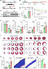
Figure 6.. Jph2 in CFs is required for fibrotic repair after myocardial infarction.
A. Breeding scheme for generating fibroblast specific Jph2 knockout (Jph2fKO) mice. B. Experimental schematic for myocardial infarction in adult Col1a2Cre and Jph2fKO mice after Tamoxifen i.p. injection. C, D. Representative (C) and quantitative (D) Western blotting for Jph2 in cardiac fibroblasts and cardiomyocytes isolated from Col1a2Cre and Jph2fKO mice after Tamoxifen injection (n=3). Gapdh was used as loading controls. E. Survival rates in Col1a2Cre and Jph2fKO mice after myocardial infarction (MI) or sham injury. N=15 for Col1a2Cre and 18 for Jph2fKO mice. F-I. Echocardiography in MI-injured or sham Col1a2Cre and Jph2fKO mice for left ventricular ejection fraction (EF%, F), end-diastolic volume (LVEDV, G), end-systolic volume (LVESV, H) and ischemic zone fraction (I) (n=5–9). J-K. Photographs (J) and quantification (K) of the area of fibrosis (blue) in transverse histological sections of Col1a2Cre and Jph2fKO mouse hearts after MI and stained with Masson’s trichrome (n=5–9). L, M. Magnification of LV scars (L) and quantified scar thickness (M) of Col1a2Cre and Jph2fKO mice heart 3 weeks after MI (n=6–7). Data are presented as mean ± SEM (D, F-I, K, and M). Statistical significance of survival was determined with the Gehan-Breslow-Wilcoxon test (E), two-way ANOVA followed by Turkey multiple comparison test (F-I and K) and unpaired t test (D and M).
Figure 7.
Figure 7.. Jph2 in CF is required for angiogenesis after myocardial infarction.
A. Vegfa, Vegfc and Vegfd gene expression in mouse CFs infected with Ad-GFP, Ad-Jph2 and Ad-shJph2, as determined by RNA-sequencing analysis. nTPM, normalized transcripts per million (n=3). B-F. Representative Western blot (B) and average data showing VEGFA (C), VEGFB (D), VEGFC (E), VEGFD (F) normalized to TBP protein levels in mouse CFs isolated from 6-week-old male TCF21iCre or TCF21iCre/shJph2 mice treated with 10 ng/mL TGFβ for 0, 1, 3, 6, 12, 24 and 48 hours. n=3 independent experiments. G-K. Sections from the infarcted and sham hearts were collected at 3 weeks after surgery and immunofluorescentlly stained for the endothelial marker CD31 (G, red) and α-smooth muscle actin (α-SMA, red) (J) relative to cardiomyocytes staining for troponin T (cTnT, green), and nuclei counterstained with DAPI (blue). CD31+ endothelial cell area as a percentage of the high-power field area (H, HPF, n=4), CD31+ vascular density (I, n=4) and α-SMA+ arterial density (K, n=4) were quantified respectively. *p<0.05; **p<0.01. Data are presented as mean ± SEM (A, C-F, H, I and K). Statistical analyses were performed using two-way ANOVA followed by Turkey multiple comparison test (A, H, I and K) and unpaired t test (C-F).
Figure 8.
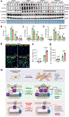
Figure 8.. Cardiac fibroblast-specific deletion of Jph2 decreases CF activation and ECM production but enhances CF proliferation after MI.
A-D. Representative Western blots (A) and quantification of FN1 (B), Col1a1 (C), and phospho-Smad3 (D) at day 3 post-MI. MI hearts were dissected into infarct, border and remote zones prior to homogenization and blotting (n=3). E-G, Immunofluorescent imaging (E) and quantification of proliferating fibroblasts (F, Ki67+/Vimentin+) and total fibroblasts (G, Vimentin+) in myocardial sections from Col1a2Cre and Jph2fKO mice 3 days post MI (n=4). Vimentin+ (green), Ki-67+ (red), nuclei (blue). n = 4 mice per group. Data are presented as mean ± SEM. Statistical analyses were performed using two-way ANOVA followed by Turkey multiple comparison test. H. Proposed model illustrating the role of junctophilin-2 (Jph2) in cardiac fibroblasts during cardiac repair under myocardial infarction stress. Top Panel: Quiescent cardiac fibroblasts are activated and differentiated into myofibroblasts in response to TGFβ signaling or myocardial infarction (MI) stress. These myofibroblasts secrete extracellular matrix (ECM) proteins and growth factors, such as VEGF, to facilitate cardiac fibrotic repair. Middle Panel: In wild-type cardiac fibroblasts, Jph2 is localized between the plasma membrane (PM) and endoplasmic reticulum (ER), where it regulates store-operated calcium entry (SOCE) by directly interacting with Stim1 and maintaining its stability. During cardiac fibroblast activation and differentiation, the expression of Jph2, along with SOCE-associated proteins like Stim1 and Orai1, is upregulated. This enhances SOCE activity, which is critical for fibroblast activation, migration, angiogenesis, and proliferation, all of which are essential for effective cardiac fibrotic repair. Bottom Panel: In the absence of Jph2 (via knockout or silencing), cardiac fibroblasts exhibit reduced SOCE activity due to the downregulation of Stim1. These fibroblasts display impaired activation, differentiation, ECM production, and angiogenesis but show increased proliferative capacity. Consequently, the loss of Jph2 disrupts cardiac fibrotic repair, leading to exacerbated cardiac damage.

References

    1. Han M, Zhou B. Role of Cardiac Fibroblasts in Cardiac Injury and Repair. Current cardiology reports. 2022;24:295–304. - PubMed
    1. Hinz B The myofibroblast: paradigm for a mechanically active cell. J Biomech. 2010;43:146–155. - PubMed
    1. Fu X, Khalil H, Kanisicak O, Boyer JG, Vagnozzi RJ, Maliken BD, Sargent MA, Prasad V, Valiente-Alandi I, Blaxall BC, et al. Specialized fibroblast differentiated states underlie scar formation in the infarcted mouse heart. The Journal of clinical investigation. 2018;128:2127–2143. - PMC - PubMed
    1. Khalil H, Kanisicak O, Vagnozzi RJ, Johansen AK, Maliken BD, Prasad V, Boyer JG, Brody MJ, Schips T, Kilian KK, et al. Cell-specific ablation of Hsp47 defines the collagen-producing cells in the injured heart. JCI Insight. 2019;4:e128722. - PMC - PubMed
    1. Wang Y, Li Q, Tao B, Angelini M, Ramadoss S, Sun B, Wang P, Krokhaleva Y, Ma F, Gu Y, et al. Fibroblasts in heart scar tissue directly regulate cardiac excitability and arrhythmogenesis. Science. 2023;381:1480–1487. - PMC - PubMed

MeSH terms