Review
. 2025 Jul 10;15(7):443.
doi: 10.3390/bios15070443.

Electrochemical Impedance Spectroscopy-Based Biosensors for Label-Free Detection of Pathogens

Affiliations
Review

Electrochemical Impedance Spectroscopy-Based Biosensors for Label-Free Detection of Pathogens

Huaiwei Zhang et al. Biosensors (Basel). .

Abstract

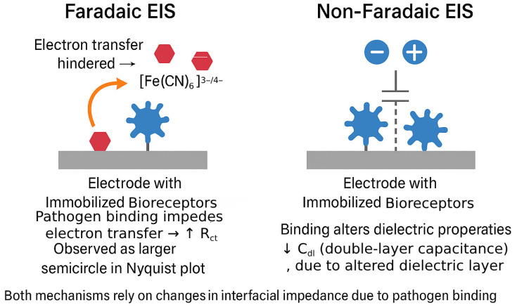
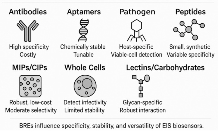
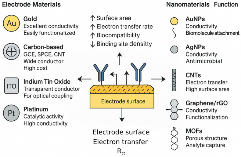
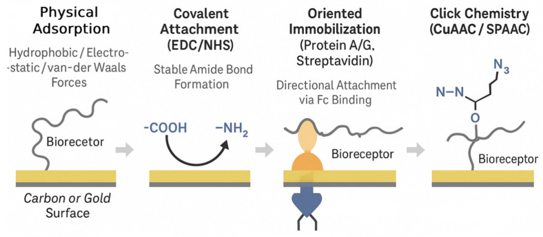
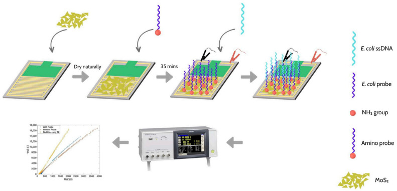
The escalating threat of infectious diseases necessitates the development of diagnostic technologies that are not only rapid and sensitive but also deployable at the point of care. Electrochemical impedance spectroscopy (EIS) has emerged as a leading technique for the label-free detection of pathogens, offering a unique combination of sensitivity, non-invasiveness, and adaptability. This review provides a comprehensive overview of the design and application of EIS-based biosensors tailored for pathogen detection, focusing on critical components such as biorecognition elements, electrode materials, nanomaterial integration, and surface immobilization strategies. Special emphasis is placed on the mechanisms of signal generation under Faradaic and non-Faradaic modes and how these underpin performance characteristics such as the limit of detection, specificity, and response time. The application spectrum spans bacterial, viral, fungal, and parasitic pathogens, with case studies highlighting detection in complex matrices such as blood, saliva, food, and environmental water. Furthermore, integration with microfluidics and point-of-care systems is explored as a pathway toward real-world deployment. Emerging strategies for multiplexed detection and the utilization of novel nanomaterials underscore the dynamic evolution of the field. Key challenges-including non-specific binding, matrix effects, the inherently low ΔRct/decade sensitivity of impedance transduction, and long-term stability-are critically evaluated alongside recent breakthroughs. This synthesis aims to support the future development of robust, scalable, and user-friendly EIS-based pathogen biosensors with the potential to transform diagnostics across healthcare, food safety, and environmental monitoring.

Keywords: biorecognition strategies; microfluidic integration; nanomaterial enhancement; non-faradaic detection; point-of-care diagnostics.


Conflict of interest statement

The authors declare no conflicts of interest.

Figures

Figure 1
Figure 1
Schematic illustration of signal generation mechanisms in label-free EIS biosensors under Faradaic and non-Faradaic modes. In the Faradaic mode (left), pathogen binding hinders the access of the redox probe ([Fe(CN)6]3−/4−) to the electrode surface, thereby increasing the Rct. In the non-Faradaic mode (right), pathogen attachment alters the dielectric environment at the electrode–electrolyte interface, reducing the Cdl. Both mechanisms lead to measurable changes in the impedance spectrum.
Figure 2
Figure 2
Visual summary of the major types of BREs employed in EIS-based pathogen biosensors. Each class—antibodies, aptamers, phages, peptides, MIPs/CIPs, whole cells, and lectins/carbohydrates—exhibits distinct physicochemical characteristics, advantages, and limitations that influence biosensor specificity, stability, cost, and viability-based detection capacity.
Figure 3
Figure 3
Schematic representation of the integration between electrode materials and nanomaterials in label-free EIS-based pathogen biosensors. Common electrode substrates, such as gold, carbon-based materials, ITO, and platinum, are shown with their key properties. Nanomaterials including AuNPs, AgNPs, CNTs, graphene derivatives, and MOFs enhance sensor performance through an improved surface area, electron transfer, biocompatibility, and functionalization capacity.
Figure 4
Figure 4
Representative strategies for immobilizing BREs on electrode surfaces in EIS-based biosensors. The illustration compares physical adsorption, covalent attachment via EDC/NHS chemistry, encapsulation in hydrogel or polymer matrices, oriented immobilization (e.g., via Protein A/G or streptavidin-biotin systems), and click chemistry (CuAAC/SPAAC). Each approach differs in stability, orientation control, and compatibility with specific biorecognition elements.
Figure 5
Figure 5
Comparative overview of EIS biosensor design strategies across bacterial, viral, fungal, and parasitic pathogens. Key features include typical biorecognition elements, common electrode materials, enhancement nanomaterials, representative detection limits, and example sample matrices.
Figure 6
Figure 6
Electrode preparation steps, including the drop-cast deposition of AuNP on bare SPEs, followed by the immobilization of FRhk-4 cells and subsequent testing with HAV [148].
Figure 7
Figure 7
Schematic illustration of one-step electrochemical aptasensor preparation [79].
Figure 8
Figure 8
The step-wise process of coating the surface of the microfabricated Au WE with SAM and the immobilization of anti-Cryptosporidium antibodies onto the microfabricated Au WE [141].
Figure 9
Figure 9
The experimental procedure of E. coli DNA detection based on hybrid MoS2 nanomaterials IDE sensors [124].

Similar articles

  • Management of urinary stones by experts in stone disease (ESD 2025).
    Papatsoris A, Geavlete B, Radavoi GD, Alameedee M, Almusafer M, Ather MH, Budia A, Cumpanas AA, Kiremi MC, Dellis A, Elhowairis M, Galán-Llopis JA, Geavlete P, Guimerà Garcia J, Isern B, Jinga V, Lopez JM, Mainez JA, Mitsogiannis I, Mora Christian J, Moussa M, Multescu R, Oguz Acar Y, Petkova K, Piñero A, Popov E, Ramos Cebrian M, Rascu S, Siener R, Sountoulides P, Stamatelou K, Syed J, Trinchieri A. Papatsoris A, et al. Arch Ital Urol Androl. 2025 Jun 30;97(2):14085. doi: 10.4081/aiua.2025.14085. Epub 2025 Jun 30. Arch Ital Urol Androl. 2025. PMID: 40583613 Review.
  • Optical and Electrochemical Biosensors for Detection of Pathogens Using Metal Nanoclusters: A Systematic Review.
    Shahrashoob M, Dehshiri M, Yousefi V, Moassesfar M, Saberi H, Molaabasi F, Zare Y, Rhee KY. Shahrashoob M, et al. Biosensors (Basel). 2025 Jul 17;15(7):460. doi: 10.3390/bios15070460. Biosensors (Basel). 2025. PMID: 40710110 Free PMC article. Review.
  • Systemic Inflammatory Response Syndrome.
    Baddam S, Burns B. Baddam S, et al. 2025 Jun 20. In: StatPearls [Internet]. Treasure Island (FL): StatPearls Publishing; 2025 Jan–. 2025 Jun 20. In: StatPearls [Internet]. Treasure Island (FL): StatPearls Publishing; 2025 Jan–. PMID: 31613449 Free Books & Documents.
  • Short-Term Memory Impairment.
    Cascella M, Al Khalili Y. Cascella M, et al. 2024 Jun 8. In: StatPearls [Internet]. Treasure Island (FL): StatPearls Publishing; 2025 Jan–. 2024 Jun 8. In: StatPearls [Internet]. Treasure Island (FL): StatPearls Publishing; 2025 Jan–. PMID: 31424720 Free Books & Documents.
  • Signs and symptoms to determine if a patient presenting in primary care or hospital outpatient settings has COVID-19.
    Struyf T, Deeks JJ, Dinnes J, Takwoingi Y, Davenport C, Leeflang MM, Spijker R, Hooft L, Emperador D, Domen J, Tans A, Janssens S, Wickramasinghe D, Lannoy V, Horn SRA, Van den Bruel A; Cochrane COVID-19 Diagnostic Test Accuracy Group. Struyf T, et al. Cochrane Database Syst Rev. 2022 May 20;5(5):CD013665. doi: 10.1002/14651858.CD013665.pub3. Cochrane Database Syst Rev. 2022. PMID: 35593186 Free PMC article.

References

    1. Cesewski E., Johnson B.N. Biosensors for the Detection of Bacterial and Viral Clinical Pathogens. Biosens. Bioelectron. 2020;159:112214. doi: 10.1016/j.bios.2020.112214. - DOI - PMC - PubMed
    1. Zhao Y.-X., Shaw P., Wen X., Dou Q., Zhang X. Recent Developments towards Portable Point-of-Care Diagnostic Devices for Pathogen Detection. Sens. Diagn. 2022;1:252–268. doi: 10.1039/d1sd00017a. - DOI
    1. Mardian Y., Kosasih H., Karyana M., Neal A., Lau C.-Y. Review of Current COVID-19 Diagnostics and Opportunities for Further Development. Front. Med. 2021;8:615099. doi: 10.3389/fmed.2021.615099. - DOI - PMC - PubMed
    1. Bhadra S., Riedel T.E., Lakhotia S., Tran N.D., Ellington A.D. High-Surety Isothermal Amplification and Detection of SARS-CoV-2. Msphere. 2021;6:e00911-20. doi: 10.1128/mSphere.00911-20. - DOI - PMC - PubMed
    1. Wang Q., Sun Y., Li Z., Yang W., Feng L. Are We Prepared for the next Pandemic: Monitor on Increasing Human and Animal H5N1 Avian Influenza Infection. Chin. Med. J. 2024;137:2776–2781. doi: 10.1097/CM9.0000000000003375. - DOI - PMC - PubMed

LinkOut - more resources