. 2025 Jul 16;15(7):458.
doi: 10.3390/bios15070458.

AI-Driven Comprehensive SERS-LFIA System: Improving Virus Automated Diagnostics Through SERS Image Recognition and Deep Learning

Affiliations

AI-Driven Comprehensive SERS-LFIA System: Improving Virus Automated Diagnostics Through SERS Image Recognition and Deep Learning

Shuai Zhao et al. Biosensors (Basel). .

Abstract

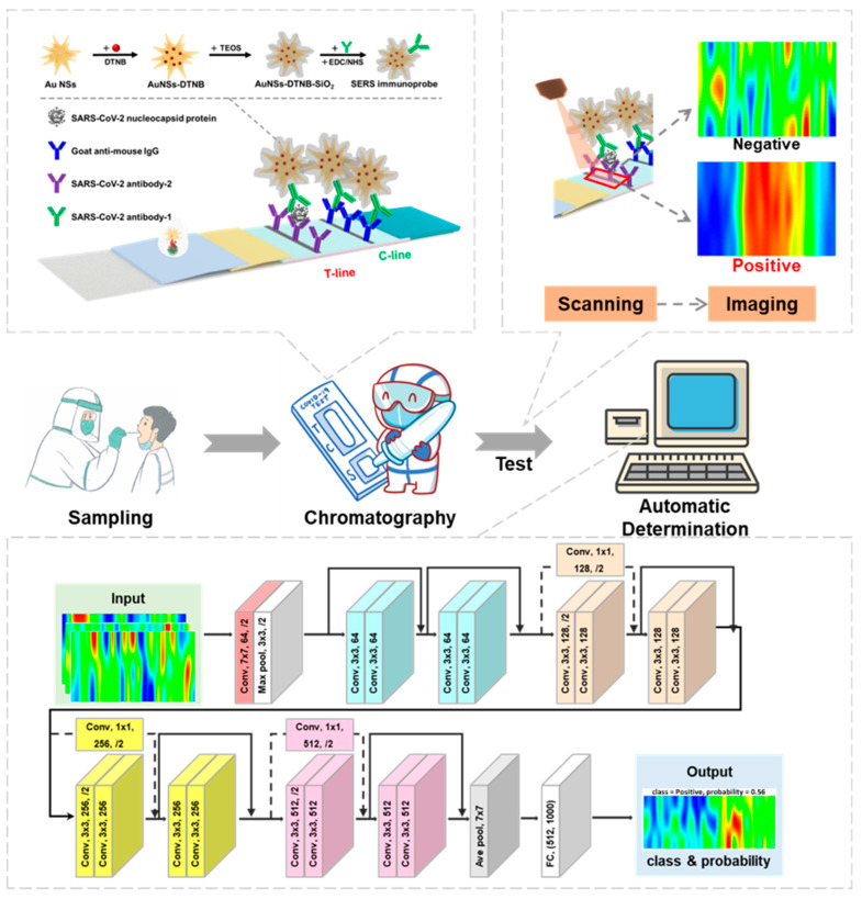
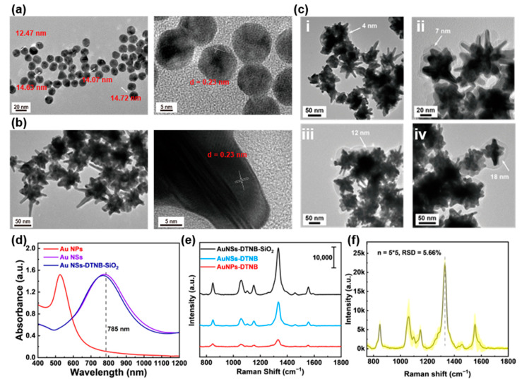
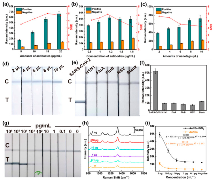
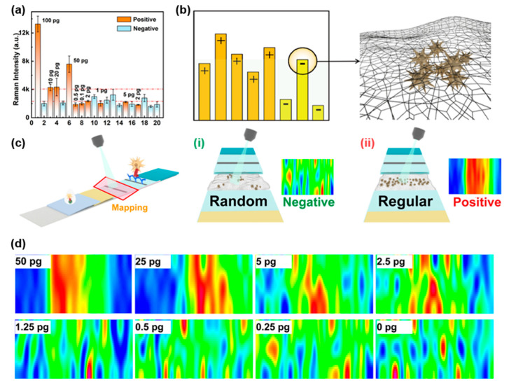
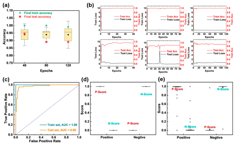
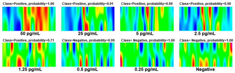
Highly infectious and pathogenic viruses seriously threaten global public health, underscoring the need for rapid and accurate diagnostic methods to effectively manage and control outbreaks. In this study, we developed a comprehensive Surface-Enhanced Raman Scattering-Lateral Flow Immunoassay (SERS-LFIA) detection system that integrates SERS scanning imaging with artificial intelligence (AI)-based result discrimination. This system was based on an ultra-sensitive SERS-LFIA strip with SiO2-Au NSs as the immunoprobe (with a theoretical limit of detection (LOD) of 1.8 pg/mL). On this basis, a negative-positive discrimination method combining SERS scanning imaging with a deep learning model (ResNet-18) was developed to analyze probe distribution patterns near the T line. The proposed machine learning method significantly reduced the interference of abnormal signals and achieved reliable detection at concentrations as low as 2.5 pg/mL, which was close to the theoretical Raman LOD. The accuracy of the proposed ResNet-18 image recognition model was 100% for the training set and 94.52% for the testing set, respectively. In summary, the proposed SERS-LFIA detection system that integrates detection, scanning, imaging, and AI automated result determination can achieve the simplification of detection process, elimination of the need for specialized personnel, reduction in test time, and improvement of diagnostic reliability, which exhibits great clinical potential and offers a robust technical foundation for detecting other highly pathogenic viruses, providing a versatile and highly sensitive detection method adaptable for future pandemic prevention.

Keywords: SARS-CoV-2; SERS-LFIA; automated detection system; deep learning; machine learning.


Conflict of interest statement

The authors declare that they have no known competing financial interests or personal relationships that could have appeared to influence the work reported in this paper.

Figures

Figure 1
Figure 1
Schematic of the SARS-CoV-2 detection and classification process including working principles of the Au NSs-DTNB-SiO2 immunoprobe-based SERS-LFIA strip; the principle of SERS scanning imaging and the architecture of the ResNet-18 image recognition model.
Figure 2
Figure 2
Characterization of Au NSs-DTNB-SiO2 immunoprobes. (a) TEM and HRTEM image of Au seeds with size 12–15 nm. (b) TEM and HRTEM image of Au NSs with size 50–100 nm. (c) TEM images of Au NSs coated with SiO2 shells of different thicknesses. (d) Absorption spectra of Au seeds, Au NSs, and SiO2-DTNB-Au NSs. (e) Raman spectra of DTNB enhanced by different nanoparticles. (f) The SERS stability of Au NSs. Note: 5 × 5 means 5 batches, with 5 measurements in each batch.
Figure 3
Figure 3
Performance evaluation of Au NSs-DTNB-SiO2 immunoprobe-based SERS-LFIA strips. (ac) Optimization of assay conditions. (d) Photographs of the Au NSs-DTNB-SiO2 immunoprobe-based SERS-LFIA strips at different immunoprobe doses. (e) Visualization results of Au NSs-DTNB-SiO2 immunoprobe-based SERS-LFIA strip specificity assays for H1N1, Influenza B virus, Influenza A virus, and HRSV recombinant proteins. (f) Intensity of the Raman spectrum at the corresponding T line in (e) around 1330 cm−1. (g) Visualization sensitivity of the Au NSs-DTNB-SiO2 immunoprobe-based SERS-LFIA strips. (h) Raman spectra of the corresponding test strips in (g). (i) Fitting curves of Raman intensity around 1330 cm−1 for different concentrations of SARS-CoV-2 NPs detected by SERS-LFIA strips based on Au NSs-DTNB-SiO2 and Au NSs-DTNB immunoprobes.
Figure 4
Figure 4
Signal analysis of SERS-LFIA strips based on a portable Raman spectroscopy. (a) SERS intensity results of 20 simulated blind samples. (b) Mechanism of false positive signals in negatives. (c) Principles of distribution-based signal discrimination: (i) negative (random probe distribution), (ii) positive (aligned distribution). (d) SERS imaging of samples at varying concentrations.
Figure 5
Figure 5
ResNet-18 model performance evaluation. (a) The 5-fold cross-validation results of the model under different epochs. (b) The loss rate and accuracy of the ResNet-18 model under different epochs of 40, 80, and 120. (c) ROC curves of the training and testing set. Scores of the (d) training and (e) testing sets.
Figure 6
Figure 6
Experimental results of simulated human sample: Nasopharyngeal swabs from healthy individuals with different concentrations of SARS-CoV-2 NPs.

Similar articles

References

    1. Peng W.Y., Wu H., Tan Y., Li M., Yang D.C., Li S. Mechanisms and treatments of myocardial injury in patients with corona virus disease 2019. Life Sci. 2020;262:118496. doi: 10.1016/j.lfs.2020.118496. - DOI - PMC - PubMed
    1. Taleghani N., Taghipour F. Diagnosis of COVID-19 for controlling the pandemic: A review of the state-of-the-art. Biosens. Bioelectron. 2021;174:112830. doi: 10.1016/j.bios.2020.112830. - DOI - PMC - PubMed
    1. Xu L.Z., Li D.Y., Ramadan S., Li Y.B., Klein N. Facile biosensors for rapid detection of COVID-19. Biosens. Bioelectron. 2020;170:112673. doi: 10.1016/j.bios.2020.112673. - DOI - PMC - PubMed
    1. Tong H.Y., Cao C.Y., You M.L., Han S., Liu Z., Xiao Y., He W.H., Liu C., Peng P., Xue Z.R., et al. Artificial intelligence-assisted colorimetric lateral flow immunoassay for sensitive and quantitative detection of COVID-19 neutralizing antibody. Biosens. Bioelectron. 2022;213:114449. doi: 10.1016/j.bios.2022.114449. - DOI - PMC - PubMed
    1. Du H., Dang X., Chen R., Li Y., Cui N., Yang H. A universal three-dimensional hydrogel electrode for electrochemical detection of SARS-CoV-2 nucleocapsid protein and hydrogen peroxide. Biosens. Bioelectron. 2024;259:116355. doi: 10.1016/j.bios.2024.116355. - DOI - PubMed

LinkOut - more resources