Review
. 2025 Jul 10;14(14):1061.
doi: 10.3390/cells14141061.

The Role of Neurohypophysial Hormones in the Endocrine and Paracrine Control of Gametogenesis in Fish

Affiliations
Review

The Role of Neurohypophysial Hormones in the Endocrine and Paracrine Control of Gametogenesis in Fish

Maya Zanardini et al. Cells. .

Abstract

Arginine vasopressin (AVP) and oxytocin (OXT) are neuropeptides traditionally recognized for their roles in the control of osmoregulation, blood pressure, lactation, and parturition in mammals. However, growing evidence suggests that AVPand OXT also regulate gonadal functions in teleost fish. Their expression in both male and female gonads, the presence of their receptors in ovaries and testes, and their interactions with steroids and other gonadal factors indicate a role in modulating gametogenesis and steroidogenesis via autocrine and paracrine mechanisms. Here, we review the current findings on AVP and OXT in teleost gonads, compared to the observed functions in mammals, emphasizing their systemic interactions within the hypothalamic-pituitary-gonadal (HPG) axis. While highlighting the roles of gonadal AVP and OXT in fish reproduction, we underscore the need for further research to unravel their complex multifactorial regulatory networks. Insights into the vasopressinergic system could enhance aquaculture practices by improving spawning success and reproductive efficiency.

Keywords: fish endocrinology; isotocin; oogenesis; paracrine regulation; spermatogenesis; steroidogenesis; vasotocin.


Conflict of interest statement

The authors declare no conflicts of interest.

Figures

Figure 1
Figure 1
(A) Multiple sequence alignment (MSA) results generated by T-Coffee for the vasopressin and oxytocin family peptides across multiple vertebrate species: human (Homo sapiens), mouse (Mus musculus), pig (Sus scrofa), zebrafish (Danio rerio), chicken (Gallus gallus), and frog (Xenopus laevis). Sequences are aligned into different regions: signal peptide, active peptide (AVP/OXT), neurophysin I or II, and copeptin. Similar colors denote the level of similarity across specific regions. Asterisks (*) mark fully conserved residues, colons (:) and periods (.) indicate strong and weak similarity, respectively. Background colors indicate conservation: warmer colors (e.g., red) show higher conservation, cooler colors (e.g., green/blue) show lower conservation. (B) Schematic representation of the precursor proteins for AVP and OXT in mammals. Each precursor consists of a signal peptide, the nonapeptide hormone (AVP/OXT), a carrier protein (neurophysin I or II), and copeptin (in the case of AVP). The dashed lines indicate cleavage sites where the precursor is processed into its active components. (C) Alignment of the core nonapeptide sequences of vasopressin/oxytocin family neuropeptides in mammals and teleost fish. The conserved cysteine residues (Cys) at positions 1 and 6 form an intramolecular disulfide bridge (S–S). Key amino acid differences between the species are highlighted in red. NCBI accession numbers in the order of sequence appearance: NP_000481.2, AAA59977.1, AAC42027.1, EDL28279.1, NP_999117.1, NP_001161061.1, NP_840078.1, NP_840076.1, NP_990516.1, XP_040527589.1, XP_018098084.1, XP_018080922.1.
Figure 2
Figure 2
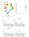
(A) UMAP representation of the cell populations in the adult ovaries of zebrafish. (B) Dot plot showing avp, avpr1ab, avpr2ab, and oxt expression levels across different cell clusters. (C) Violin plots representing gene expression. Each violin plot depicts the expression distribution of avp, avpr1ab, avpr2ab, and oxt across different cell types. Cell types include follicle cells, stromal cells, germline stem cells (GSCs), cells entering meiosis (meio_entry), transit-amplifying cells (trans_amp), natural killer-like cells (NK-like), post-meiotic germ cells (post_meio), early oocytes (early_oo and early_oo2), vasculature-associated cells, meiotic oocytes (meio), theca cells, macrophages, neutrophils, and a small group of unidentified cells.
Figure 3
Figure 3
(A) UMAP representation of 10 different cell populations in adult zebrafish testes. (B) Dot plot showing expression levels of avp, avpr1aa, avpr1ab, avpr2a, oxt, oxtra, and oxtrb across different cell clusters. (C) Violin plots representing gene expression. Each violin plot depicts the expression distribution of avp, avp receptors, oxt, and oxt receptors across different cell types. Cell types include type A undifferentiated spermatogonia (Aund), type A differentiated spermatogonia (Adiff), early type B spermatogonia (typeB), late type B spermatogonia (late typeB), germ cells that enter meiosis I (Spermatocytes1), germ cells that enter meiosis II (Spermatocytes2), haploid cells before the final maturation (round spermatids), fully mature haploid cells (elongated spermatids), Leydig cells (Leydig), Sertoli cells (Sertoli), and a small group of immune and peritubular myoid cells (Immune/myoid).
Figure 4
Figure 4
Schematic illustration of the factors involved in the autocrine/paracrine regulation of ovarian functions and oocyte development in fish, including vasotocin (avp). Oocyte classification follows the zebrafish nomenclature: I—primary-growth oocyte; II—secondary-growth oocyte; III—previtellogenic oocyte; IV—vitellogenesis; V—mature oocyte undergoing GVBD. Abbreviations: E2: estradiol-17β; DHP: 17α,20β-dihydroxy-4-pregnen-3-one; bmp15: bone morphogenic protein 15; amh: anti-Müllerian hormone; igf3: insulin-like growth factor 3; avp: arginine-vasotocin; avpr1: vasotocin receptor type 1; avpr2: vasotocin receptor type 2; PGF: prostaglandin F2α; PGE2: prostaglandin E2; GnIH: gonadotropin-inhibitory hormone; GnRH: gonadotropin-releasing hormone; H2O: water. Each factor is depicted in a unique color to facilitate clear identification.
Figure 5
Figure 5
Schematic representation of the local role of vasotocin (avp) on zebrafish spermatogenesis. Germ cell development includes undifferentiated spermatogonia or stem cells (Aund*, the asterisk marks Aund stem/progenitor cells), type A undifferentiated non-stem cells (Aund), type A differentiated cells (Adiff), type B cells (SpgB), spermatocytes, spermatids, and spermatozoa. Abbreviations: gdnf: glial cell line-derived neurotrophic factor; amh: anti-Müllerian hormone; igf3: insulin-like growth factor 3; inls3: insulin-like peptide 3; avp: arginine-vasotocin; PGE2: prostaglandin E2; Wnt5a: wingless-type MMTV integration site family member 5A; RA: retinoic acid. Each factor is depicted in a unique color to facilitate clear identification.
Figure 6
Figure 6
Schematic overview of neuroendocrine and paracrine/autocrine regulation of reproduction in female (top) and male (bottom) fish. The diagram illustrates the integration of neurosecretory hormones and classical endocrine signals from the anterior and posterior pituitary, highlighting the roles of AVP and OXT in both central and local (paracrine/autocrine) regulation. Key reproductive hormones and factors are shown, including gonadotropin-releasing hormone (GnRH), gonadotropin-inhibitory hormone (GnIH), gamma-aminobutyric acid (GABA), neuropeptide Y (NPY), luteinizing hormone (LH), follicle-stimulating hormone (FSH), estradiol-17β (E2), progesterone (P4), 17α-hydroxyprogesterone (17-OHP4), 17α,20β-dihydroxy-4-pregnen-3-one (DHP), testosterone (T), 11-ketotestosterone (11-KT), insulin-like growth factor 3 (igf3), epidermal growth factor (EGF), bone morphogenetic protein 15 (bmp15), growth differentiation factor 9 (GDF9), anti-Müllerian hormone (amh), glial cell line-derived neurotrophic factor (gdnf), Wnt family member 5A (wnt5a), insulin-like peptide 3 (insl3), and retinoic acid (RA).

Similar articles

References

    1. Kelly A.M., Goodson J.L. Behavioral relevance of species-specific vasotocin anatomy in gregarious finches. Front. Neurosci. 2013;7:242. doi: 10.3389/fnins.2013.00242. - DOI - PMC - PubMed
    1. Goodson J.L. Nonapeptides and the evolutionary patterning of sociality. Prog. Brain Res. 2008;170:3–15. doi: 10.1016/S0079-6123(08)00401-9. - DOI - PMC - PubMed
    1. Santangelo N., Bass A.H. Individual behavioral and neuronal phenotypes for arginine vasotocin mediated courtship and aggression in a territorial teleost. Brain Behav. Evol. 2010;75:282–291. doi: 10.1159/000316867. - DOI - PubMed
    1. Godwin J., Thompson R. Nonapeptides and Social Behavior in Fishes. Horm. Behav. 2012;61:230–238. doi: 10.1016/j.yhbeh.2011.12.016. - DOI - PubMed
    1. Cardoso S.C., Paitio J.R., Oliveira R.F., Bshary R., Soares M.C. Arginine vasotocin reduces levels of cooperative behaviour in a cleaner fish. Physiol. Behav. 2015;139:314–320. doi: 10.1016/j.physbeh.2014.11.052. - DOI - PubMed

LinkOut - more resources