. 2025 Jul 11;14(14):1062.
doi: 10.3390/cells14141062.

Standardized Workflow and Analytical Validation of Cell-Free DNA Extraction for Liquid Biopsy Using a Magnetic Bead-Based Cartridge System

Affiliations

Standardized Workflow and Analytical Validation of Cell-Free DNA Extraction for Liquid Biopsy Using a Magnetic Bead-Based Cartridge System

Shivaprasad H Sathyanarayana et al. Cells. .

Abstract

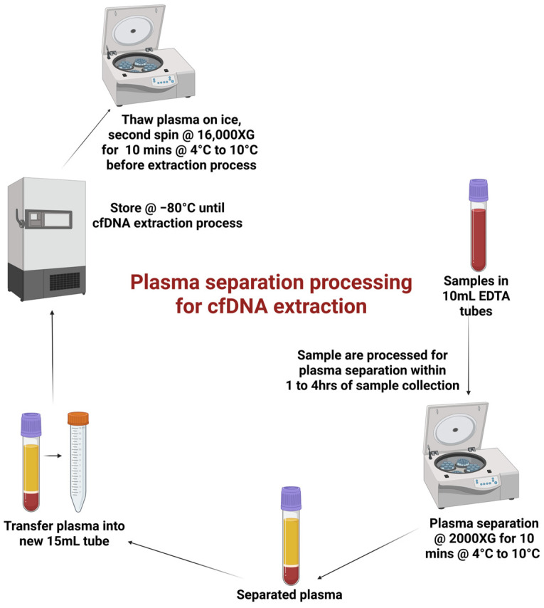
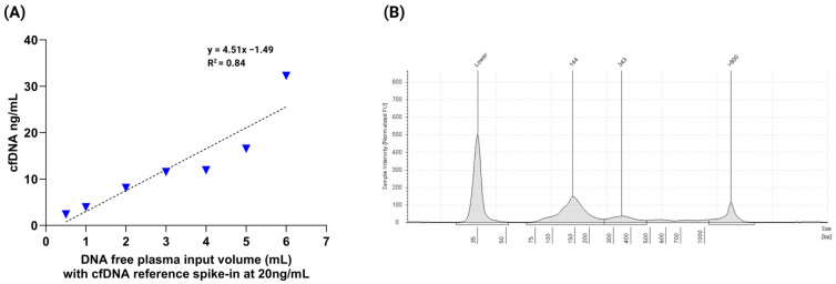
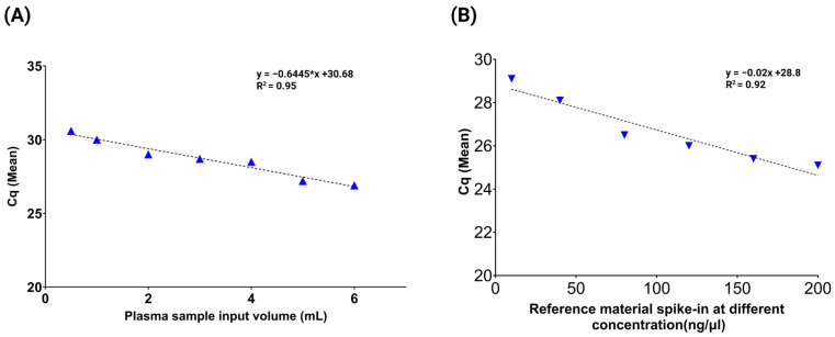
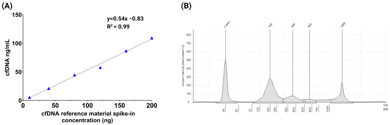
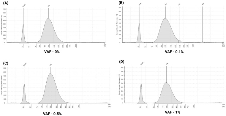
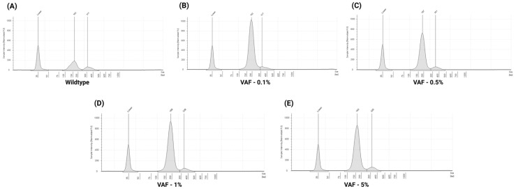
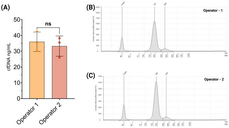
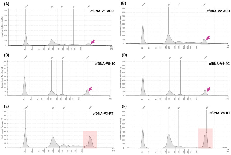
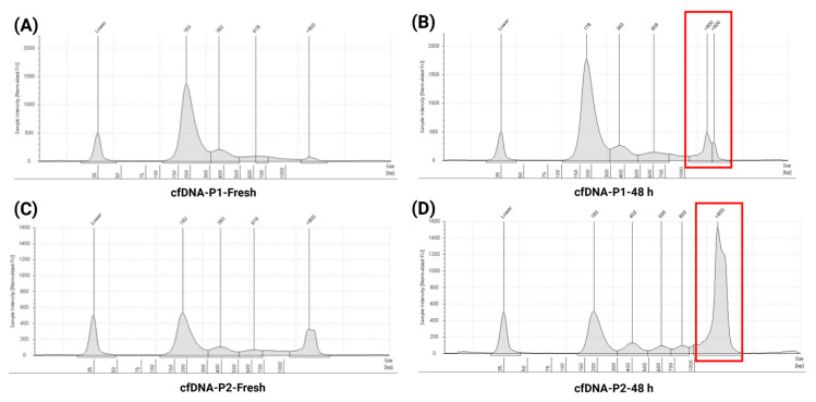
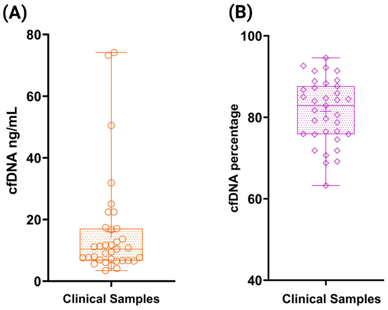
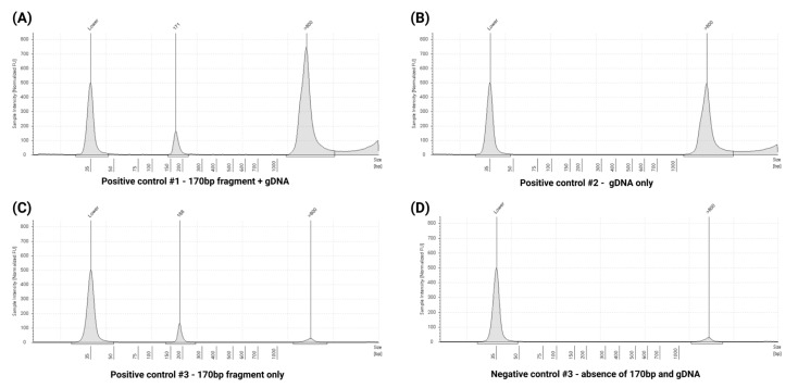
Circulating cell-free DNA (cfDNA) is an important biomarker for various cancer types, enabling a non-invasive testing approach. However, pre-analytical variables, including sample collection, tube type, processing conditions, and extraction methods, can significantly impact the yield, integrity, and overall quality of cfDNA. This study presents a comprehensive analytical validation of a magnetic bead-based, high-throughput cfDNA extraction system, with a focus on assessing its efficiency, reproducibility, and compatibility with downstream molecular applications. The validation was performed using a range of sample types: synthetic cfDNA spiked into DNA-free plasma, multi-analyte ctDNA plasma controls, Seraseq ctDNA reference material in a plasma-like matrix, extraction specificity controls, residual clinical specimen from patients, and samples from healthy individuals stored at room temperature or 4 °C for up to 48 h to assess stability. Extracted cfDNA was analyzed for concentration, percentage, and fragment size, using the Agilent TapeStation. Variant detection was evaluated using a next-generation sequencing (NGS) assay on the Seraseq ctDNA reference material. The results demonstrated high cfDNA recovery rates, consistent fragment size distribution (predominantly mononucleosomal and dinucleosomal), minimal genomic DNA (gDNA) contamination, and strong concordance between detected and expected variants in reference materials. The workflow also showed robust performance under different study parameters, variable sample conditions, including sample stability and integrity. Together, these findings confirm the efficiency and reliability of the evaluated cfDNA extraction system and underscore the importance of standardized pre-analytical workflows for the successful implementation of liquid biopsy for early cancer detection, therapeutic monitoring, and improved patient outcomes.

Keywords: TapeStation; cancer detection; cell-free DNA; cfDNA extraction; liquid biopsy; reference materials.


Conflict of interest statement

The authors declare no conflicts of interest.

Figures

Figure 1
Figure 1
Extraction workflow—optimized plasma separation workflow for cfDNA extraction.
Figure 2
Figure 2
Linearity and fragment profile of cfDNA extraction using cfDNA reference material. (A) Linearity analysis of the cfDNA extraction method performed using nRichDX reference material spiked at a fixed concentration (20 ng/mL) into varying plasma input volumes (0.5–6 mL). A strong linear correlation (R2 ≥ 0.84) was observed between input volume and recovered cfDNA concentration, indicating consistent extraction efficiency across sample volumes. (B) Representative Agilent TapeStation electropherogram of the extracted cfDNA, displaying distinct at ~144 bp (mononucleosomal) and ~343 bp (dinucleosomal), with minimal gDNA traces observed at fragment sizes > 800 bp.
Figure 3
Figure 3
cfDNA reference material extraction recovery using qPCR targeting the KRAS p.G12V variant. (A) qPCR analysis of cfDNA extracted from plasma samples spiked with 20 ng/mL of KRAS p.G12V reference material across varying plasma input volumes. The results demonstrated efficient and consistent cfDNA recovery with a strong linear correlation (R2 ≥ 0.95), indicating volume-independent extraction performance. (B) cfDNA recovery efficiency was further assessed using a range of cfDNA spike-in concentrations. The qPCR data showed a robust linear relationship (R2 ≥ 0.92), confirming the quantitative accuracy and reliability of the extraction method across a dynamic input range.
Figure 4
Figure 4
Data representing varying input concentrations. (A) Linearity assessment using the cfDNA reference material spiked into 2 mL of plasma at input concentrations ranging from 10 ng to 200 ng. The extraction demonstrated excellent linearity and high recovery efficiency with a strong correlation (R2 ≥ 0.99) using the Revolution workflow. (B) Representative TapeStation electropherogram showing cfDNA fragment size distribution, with distinct peaks at approximately149 bp (mononucleosomal), 358 bp (dinucleosomal), and 563 bp (trinucleosomal).
Figure 5
Figure 5
Representative TapeStation electropherogram profiles of multi-analyte ctDNA plasma control: electropherogram peaks illustrating cfDNA fragment size distributions across different VAF: (A) 185 bp (VAF—0%), (B) 190 bp (VAF—0.1%), (C) 183 bp (VAF—0.5%), and (D) 184 bp (VAF—1%). All profiles show well-defined cfDNA peaks with no gDNA contamination, as indicated by the absence or minimal signal above 800 bp.
Figure 6
Figure 6
Representative TapeStation electropherogram profiles of ctDNA complete reference material: electropherogram peaks illustrating cfDNA fragment size distributions across different VAF: (A) 185 bp—mononucleosomal, 311—dinucleosomal (Wildtype), (B) 193 bp—mononucleosomal, 317—dinucleosomal (VAF—0.1%), (C) 187 bp—monomer, 341—dinucleosomal (VAF—0.5%), (D) 189 bp—mononucleosomal, 338—dinucleosomal (VAF—1%), and (E) 163 bp—mononucleosomal, 320—dinucleosomal (VAF—5%). Notably, no gDNA contamination was observed, as indicated by the absence of high-intensity peaks above 800 bp.
Figure 7
Figure 7
Precision and reproducibility of cfDNA extraction. (A) Total cfDNA yield obtained by two independent operators using the same extraction protocol and sample input, demonstrating consistent recovery and reproducibility. (B,C) Representative TapeStation electropherograms from each operator show highly similar cfDNA fragment profiles, with dominant peaks at ~180–190 bp (mononucleosomal) and ~340 bp (dinucleosomal), indicating reproducible fragment recovery and minimal inter-operator variability.
Figure 8
Figure 8
Impact of room temperature and 4°C storage on cfDNA stability in blood samples over a 48 h period compared to fresh samples. (A,B) TapeStation electropherograms of cfDNA extracted from freshly collected blood samples in ACD tubes, showing characteristic fragment peaks and minimal gDNA contamination. (C,D) Electropherograms from samples stored at 4 °C for up to 48 h demonstrating preserved cfDNA integrity with minimal gDNA presence compared to fresh samples. (E,F) Electropherograms from clinical samples stored at RT for up to 48 h, showing distinct cfDNA fragment peaks around ~180–190 bp (mononucleosomal), ~350–400 bp (dinucleosomal), along with significantly increased gDNA contamination (>800 bp). High-molecular-weight gDNA levels are indicated by arrows in panels (AD), and highlighted in red boxes in panels (E,F), illustrating significant amount of gDNA due to the adverse impact of RT storage on cfDNA purity and integrity.
Figure 9
Figure 9
Assessment of cfDNA stability in blood samples stored at RT for up to 48 h compared to freshly processed samples. (A,C) TapeStation electropherograms of cfDNA extracted from freshly processed clinical blood samples, showing distinct cfDNA fragment peaks with minimal gDNA contamination. (B,D) Electropherograms of cfDNA extracted from matched clinical samples stored at RT for up to 48 h, demonstrating characteristic cfDNA fragment peaks around ~180 bp (mononucleosomal), ~370–380 bp (dinucleosomal), and ~600 bp (trinucleosomal), accompanied by marked gDNA contamination (>800 bp), as highlighted in the red boxes. These findings underscore the adverse impact of prolonged RT storage on cfDNA integrity and purity, and highlight the importance of timely plasma processing to minimize pre-analytical variability.
Figure 10
Figure 10
cfDNA extraction and quantification from clinical samples. (A) Concentration of extracted cfDNA from clinical samples quantified using the Agilent TapeStation and expressed in ng/mL. (B) Percentage of cfDNA in each patient sample as determined by TapeStation analysis.
Figure 11
Figure 11
TapeStation electropherogram profiles of extraction specificity controls used for quality control assessments. (A) Positive control 1: Mixed sample containing both gDNA and 170 bp cfDNA fragments, demonstrating distinct peaks for each component. (B) Positive control 2: Sample containing gDNA only, showing a high-molecular-weight peak with no cfDNA signal. (C) Positive control 3: Sample containing 170 bp cfDNA fragments with minimal gDNA contamination, indicating successful cfDNA isolation. (D) Negative control: No detectable peaks, confirming the absence of contamination or non-specific amplification.

Similar articles

References

    1. Van Der Vaart M., Pretorius P.J. The Origin of Circulating Free DNA. Clin. Chem. 2007;53:2215. doi: 10.1373/clinchem.2007.092734. - DOI - PubMed
    1. Qin Z., Ljubimov V.A., Zhou C., Tong Y., Liang J. Cell-Free Circulating Tumor DNA in Cancer. Chin. J. Cancer. 2016;35:36. doi: 10.1186/s40880-016-0092-4. - DOI - PMC - PubMed
    1. Volckmar A., Sültmann H., Riediger A., Fioretos T., Schirmacher P., Endris V., Stenzinger A., Dietz S. A Field Guide for Cancer Diagnostics Using Cell-free DNA: From Principles to Practice and Clinical Applications. Genes. Chromosomes Cancer. 2018;57:123–139. doi: 10.1002/gcc.22517. - DOI - PubMed
    1. Udomruk S., Orrapin S., Pruksakorn D., Chaiyawat P. Size Distribution of Cell-Free DNA in Oncology. Crit. Rev. Oncol./Hematol. 2021;166:103455. doi: 10.1016/j.critrevonc.2021.103455. - DOI - PubMed
    1. Vellanki P.J., Ghosh S., Pathak A., Fusco M.J., Bloomquist E.W., Tang S., Singh H., Philip R., Pazdur R., Beaver J.A. Regulatory Implications of ctDNA in Immuno-Oncology for Solid Tumors. J. Immunother. Cancer. 2023;11:e005344. doi: 10.1136/jitc-2022-005344. - DOI - PMC - PubMed

Publication types

LinkOut - more resources