. 2025 Jul 11;14(14):1067.
doi: 10.3390/cells14141067.

Assessment of Feasibility of the M2 Macrophage-Based Adoptive Gene Transfer Strategy for Osteoarthritis with a Mouse Model

Affiliations

Assessment of Feasibility of the M2 Macrophage-Based Adoptive Gene Transfer Strategy for Osteoarthritis with a Mouse Model

Matilda H-C Sheng et al. Cells. .

Abstract

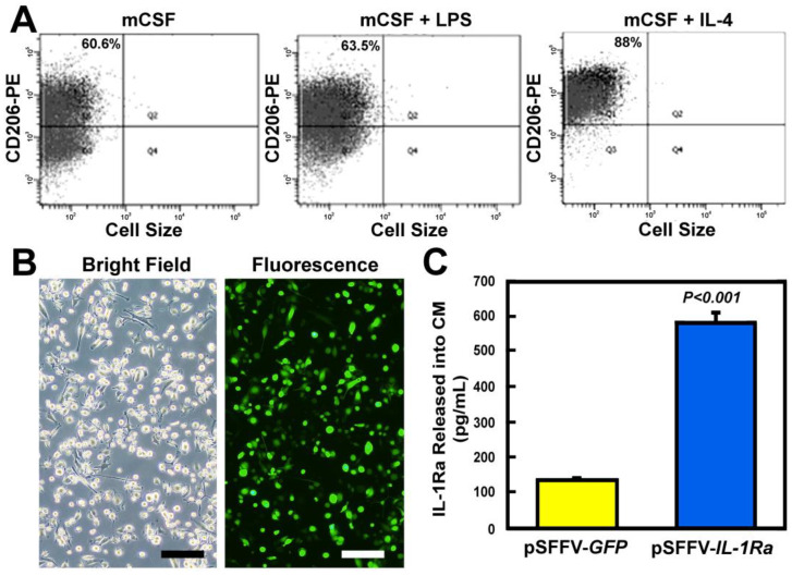
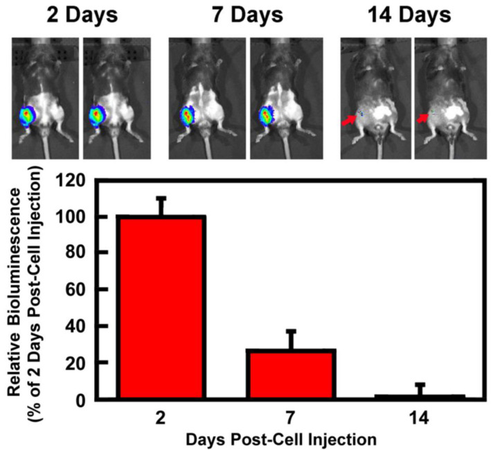
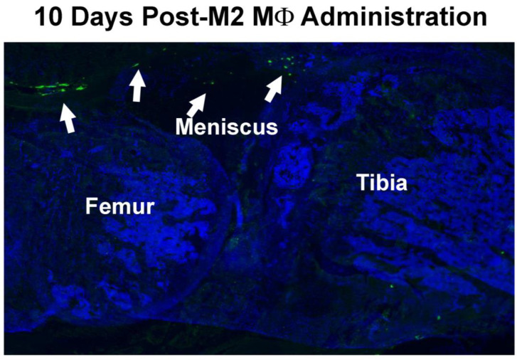
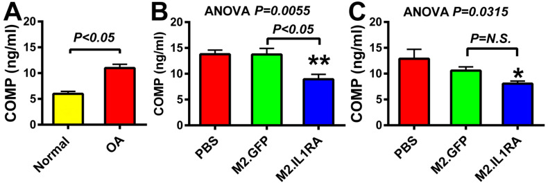
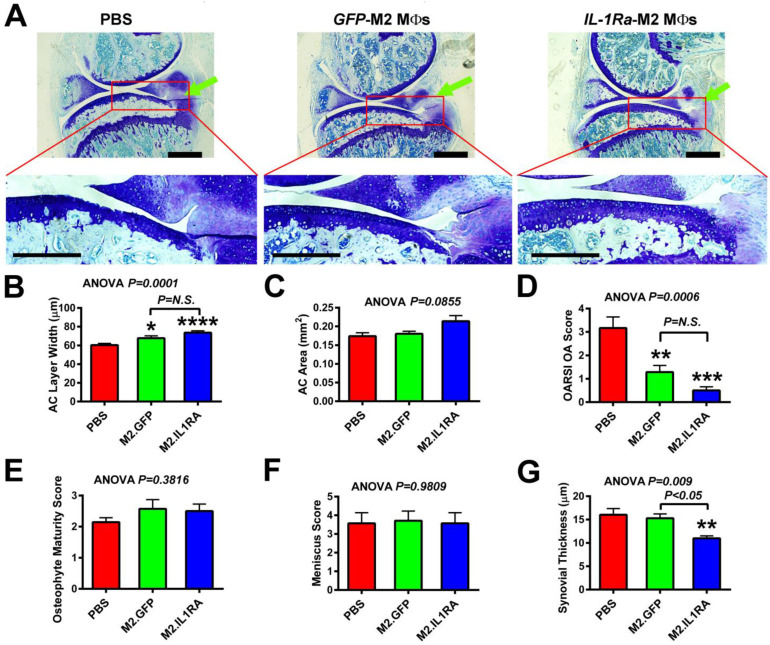
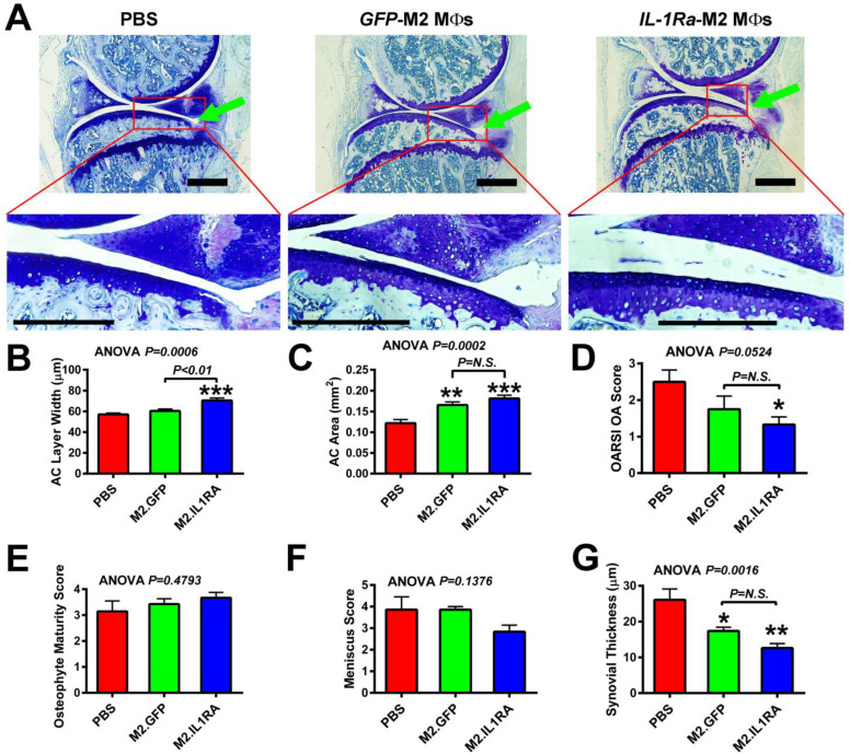
Current osteoarthritis (OA) therapies fail to yield long-term clinical benefits, due in part to the lack of a mechanism for the targeted and confined delivery of therapeutics to OA joints. This study evaluates if M2 macrophages are effective cell vehicles for the targeted and confined delivery of therapeutic genes to OA joints. CT bioluminescence in vivo cell tracing and fluorescent microscopy reveal that intraarticularly injected M2 macrophages were recruited to and retained at inflamed synovia. The feasibility of an M2 macrophage-based adoptive gene transfer strategy for OA was assessed using IL-1Ra as the therapeutic gene in a mouse tibial plateau injury model. Mouse M2 macrophages were transduced with lentiviral vectors expressing IL-1Ra or GFP. The transduced macrophages were intraarticularly injected into injured joints at 7 days post-injury and OA progression was monitored with plasma COMP and histology at 4 weeks. The IL-1Ra-expressing M2 macrophage treatment reduced plasma COMP, increased the area and width of the articular cartilage layer, decreased synovium thickness, and reduced the OARSI OA score without affecting the osteophyte maturity and meniscus scores when compared to the GFP-expressing M2 macrophage-treated or PBS-treated controls. When the treatment was given at 5 weeks post-injury, at which time OA should have developed, the IL-1Ra-M2 macrophage treatment also reduced plasma COMP, had a greater articular cartilage area and width, decreased synovial thickness, and reduced the OARSI OA score without an effect on the meniscus and osteophyte maturity scores at 8 weeks post-injury. In conclusion, the IL-1Ra-M2 macrophage treatment, given before or after OA was developed, delayed OA progression, indicating that the M2 macrophage-based adoptive gene transfer strategy for OA is tenable.

Keywords: IL-1Ra; adoptive gene transfer; inflammation; macrophages; osteoarthritis; treatment.


Conflict of interest statement

All authors (Sheng, M.H.-C., Baylink, D.J., Rundle, C.H. and Lau, K.-H.W.) state that they have no conflicts of interest to declare. The views, opinions, and/or findings contained in this report are those of the authors and should not be construed as an official position, policy, or decision of the US Army, the US Department of Veterans’ Affairs, or the United States government, unless so designated by other documentation. The funders had no role in study design, data collection and analysis, decision to publish, or preparation of the manuscript.

Figures

Figure 1
Figure 1
Levels of IL-1Ra protein secreted into CM by mCSF/IL-4-expanded pSFFV-IL-1Ra-transduced M2 MΦs in cultures. (A) Florescence-assisted cell sorting (FACS) analysis of murine M2 MΦs for CD206, a biomarker specific for M2 MΦs, after expansion with mCSF alone (left), expansion and M1 polarization with the mCSF and LPS co-treatment (middle), and expansion and M2 polarization with the m-CSF and IL-14 co-treatment (right). [Each expanded and polarized cell population was also sorted for CD11b and F4/80, and each was >97 and >98% positive for CD11b and F4/80, respectively, indicating that there were indeed MΦs.] (B) A representative bright-field (left) and fluorescent (right) photomicrograph of GFP expression in a representative culture of IL-1Ra/GFP-expressing murine M2 MΦs. The transduction efficiency in this experiment, judged by GFP expression, was estimated to be ~70%. Scale bars = 20 µm. (C) The CM of the transduced cells after 72 h in culture was collected and the relative IL-1Ra protein level in CM was measured by ELISA. Results are shown as mean ± SEM with n = 4 per group. Statistical significance of the difference between the two groups was determined with two-tailed Student’s t-test.
Figure 2
Figure 2
Temporal and spatial tracing of the luciferase-expressing M2 MΦs in the injured knee joint in vivo after intraarticular injection. Intraarticular tibial plateau injury was created on the right knee joint of 10-week-old C57BL/6J mice. At 7 days post-injury, Luc-expressing M2 MΦs were injected intraarticularly. At 2, 7, and 14 days after the cell injection, D-luciferin was injected i.p. into each mouse. The tracking of the injected Luc-expressing M2 MΦs was performed with Perkin-Elmer IVIS CT bioluminescence in vivo imaging system. Top is the CT bioluminescence image of two representative mice at each time point. Bottom shows the relative levels of bioluminescence emitted, as a percentage of bioluminescence emitted on day 2. Results are shown as mean ± SEM (n = 5).
Figure 3
Figure 3
Recruitment and retention of intraarticularly injected M2 MΦs in the inflamed synovial membrane. A representative fluorescent photomicrograph of the location of GFP-expressing M2 MΦs in the injected joint. GFP-expressing M2 MΦs (isolated from GFP transgenic mice) were injected intraarticularly into the injured joint on day 4 post-injury and sagittal sections of the injured knee joint were examined by fluorescent microscopy 10 days later (i.e., 17 days post-injury).
Figure 4
Figure 4
Plasma levels of COMP in mice treated with M2 MΦs expressing IL1Ra at 1 or 5 weeks post-tibial plateau injury. (A) Plasma COMP levels of normal healthy mice (left) or untreated OA mice at two weeks post-tibial plateau injury. Statistical significance was determined by two-tailed Student’s t-test. (B) Plasma COMP levels at 4 weeks post-injury in mice receiving a single injection of IL-1Ra- or GFP-expressing M2 MΦs at 1 week post-injury. (C) Plasma COMP levels at 8 weeks post-injury in mice receiving a single injection of IL-1Ra- or GFP-expressing M2 MΦs at 5 weeks post-injury. N = 6–7 mice per test group. Statistical significance in (B,C) was determined by one-way ANOVA followed by Tukey’s post hoc test. * p <0.05 and ** p < 0.01 compared to the PBS control group. P = N.S. (not significant), when p > 0.05.
Figure 5
Figure 5
Effects of a single intraarticular injection of M2 MΦs expressing IL1Ra (M2.IL1RA) or GFP (M2.GFP), or PBS alone, at 1 week after the tibial plateau injury on OA progression. Tibial plateau injury was created on the right knee. The genetically modified M2 MΦs or PBS were injected intraarticularly at 1 week after injury. OA progression was monitored at 4 weeks post-injury by histology on sagittal thin sections (from the medial side). (A) Top, toluidine blue staining of cartilage of a representative injured joint of each treatment group. The green arrow indicates the site of compression loading on tibial plateau. Bottom, higher magnification of tibial plateau articular cartilage layer in each red box of the indicated knee joint. Scale bars = 250 µm. (B) Average articular cartilage (AC) layer width of tibial plateau; (C) average articular cartilage (AC) layer area; (D) OARSI OA score; (E) osteophyte maturity score; (F) meniscus score; and (G) average thickness of synovial layers. Results are shown as mean ± SEM, N = 6–7 per group. Statistical significance was determined by one-way ANOVA followed by Tukey’s post hoc test. * p < 0.05, ** p < 0.01, *** p < 0.001, and **** p < 0.0001, when compared with PBS (“no treatment”) group. P = N.S. (not significant), when p > 0.05.
Figure 6
Figure 6
Effects of a single intraarticular injection of M2 MΦs expressing IL1Ra (M2.IL1RA) or GFP (M2.GFP), or PBS alone, at 5 weeks after the tibial plateau injury on OA progression. Tibial plateau injury was created on the right knee. Genetically modified M2 MΦs were injected at 5 weeks after injury, at which time OA should have been developed [39,45,46]. OA progression was determined 3 weeks later by histology on sagittal thin sections (from the medial side). (A) Top, toluidine blue staining of cartilage of a representative injured joint of each treatment group. Green arrow indicates the site of compression loading on the tibial plateau. Bottom, higher magnification of tibial plateau articular cartilage layer in each red box of the indicated knee joint. Scale bars = 250 µm. (B) Average articular cartilage (AC) layer width on the tibia of injured joints; (C) average articular cartilage (AC) layer area; (D) OARSI OA score; (E) osteophyte maturity score; (F) meniscus score; and (G) average thickness of the synovial layer. In each panel, results are shown as mean ± SEM, N = 6–7 per group. Statistical significance was determined by one-way ANOVA followed by Tukey’s post hoc test. * p < 0.05, ** p < 0.01, and *** p < 0.001, when compared with PBS (“no treatment”) group. P = N.S. (not significant), when p > 0.05.

Similar articles

References

    1. Martin J.A., Buckwalter J.A. Post-traumatic osteoarthritis: The role of stress induced chondrocyte damage. Biorheology. 2006;43:517–521. doi: 10.1177/0006355X2006043003004034. - DOI - PubMed
    1. Brown T.D., Johnston R.C., Saltzman C.L., Marsh J.L., Buckwalter J.A. Posttraumatic osteoarthritis: A first estimate of incidence, prevalence, and burden of disease. J. Orthop. Trauma. 2006;20:739–744. doi: 10.1097/01.bot.0000246468.80635.ef. - DOI - PubMed
    1. Sharma L. Osteoarthritis of the Knee. N. Eng. J. Med. 2021;384:51–59. doi: 10.1056/NEJMcp1903768. - DOI - PubMed
    1. Krakowski P., Rejniak A., Sobczyk J., Karpinski R. Cartilage Integrity: A Review of Mechanical and Frictional Properties and Repair Approaches in Osteoarthritis. Healthcare. 2024;12:1648. doi: 10.3390/healthcare12161648. - DOI - PMC - PubMed
    1. Leifer V.P., Katz J.N., Losina E. The burden of OA-health services and economics. Osteoarthr. Cartil. 2022;30:10–16. doi: 10.1016/j.joca.2021.05.007. - DOI - PMC - PubMed

Substances

LinkOut - more resources