. 2025 Jul 13;14(14):1070.
doi: 10.3390/cells14141070.

Regulation of Hindbrain Vascular Development by rps20 in Zebrafish

Affiliations

Regulation of Hindbrain Vascular Development by rps20 in Zebrafish

Xinyu Shen et al. Cells. .

Abstract

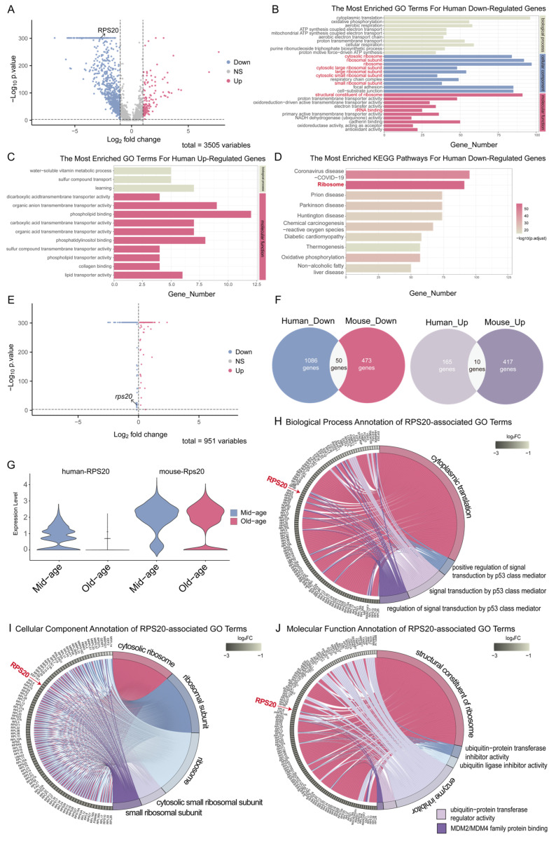
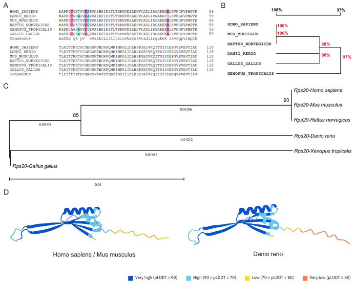
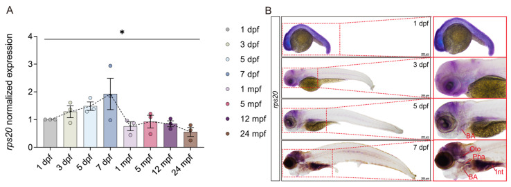
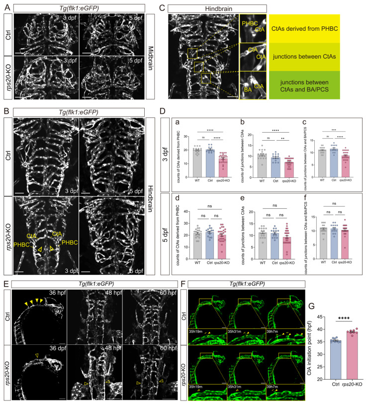
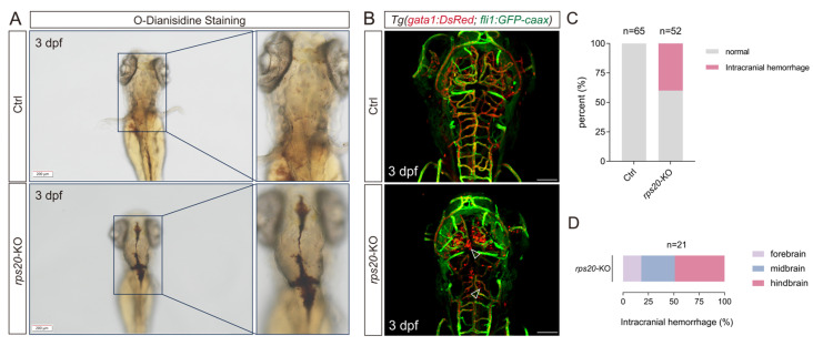
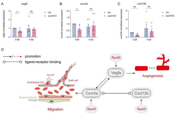
During aging, the brain vasculature undergoes significant deterioration characterized by increased arterial tortuosity, compromised blood-brain barrier integrity, and reduced cerebral blood flow, all of which contribute to various neurological disorders. Thus, understanding the mechanisms underlying aging-related cerebrovascular defects is critical for developing strategies to alleviate aging-associated neurological diseases. In this study, we investigated the role of aging-related genes in brain vascular development using zebrafish as an in vivo model. By thoroughly analyzing scRNA-seq datasets of mid- and old-aged brain vascular endothelial cells (human/mouse), we found ribosomal protein S20 (rps20) significantly down-regulated during aging. qPCR analysis and whole-mount in situ hybridization validated a high expression of rps20 during early zebrafish development, which progressively decreased in adult and aged zebrafish brains. Functional studies using the CRISPR/Cas9-mediated knockout of rps20 revealed an impaired growth of central arteries in the hindbrain and a marked increased intracranial hemorrhage incidence. Mechanistically, qPCR analysis demonstrated a significant downregulation of vegfa, cxcl12b, and cxcr4a, key signaling molecules required for hindbrain vascular development, in rps20-deficient embryos. In conclusion, our findings demonstrate that rps20 is essential for proper brain vascular development and the maintenance of vascular homeostasis in zebrafish, revealing a novel mechanism by which aging-related genes regulate brain vascular development. This study provides new insights that may aid in understanding and treating aging-associated vascular malformations and neurological pathologies.

Keywords: aging; brain vasculature; rps20; vascular permeability; zebrafish.


Conflict of interest statement

The authors declare no conflicts of interest.

Figures

Figure 1
Figure 1
Cross-species bioinformatics identified conserved aging signatures in cerebrovascular endothelial cells. (A) Volcano plots showing the differentially expressed genes in mid- and old-aged human cerebrovascular endothelial cells; (B) GO analysis of down-regulated genes in aged human cerebrovascular endothelial cells; (C) GO analysis of up-regulated genes in aged human cerebrovascular endothelial cells; (D) KEGG analysis of down-regulated genes in aged human cerebrovascular endothelial cells; (E) Volcano plots showing the differentially expressed genes in mid- and old-aged mouse cerebrovascular endothelial cells; (F) Wayne diagrams showing the overlapped up and down-regulated genes in aged human and mouse cerebrovascular endothelial cells; (G) Expression of rps20 in human and mouse cerebrovascular endothelial cells in mid-aged and old-aged adults; (HJ) Genes and signaling pathways involved in the rps20 participated GO terms.
Figure 2
Figure 2
Evolutionary conservation of RPS20 protein in vertebrates. (A) Multiple sequence alignment analysis of RPS20 protein sequences from six representative species. Red sequences indicate homology level ≥75% and blue sequences indicate homology level ≥50%; (B) Homologous tree constructed based on the multiple sequence alignment of RPS20; (C) Phylogenetic tree of the RPS20 protein showing the high evolutionary conservation of RPS20 protein; (D) 3D structures of human, mouse, and zebrafish RPS20 proteins predicted by AlphaFold. pLDDT (per-residue confidence score) indicating residue-level confidence of AlphaFold-predicted structures.
Figure 3
Figure 3
Spatiotemporal expression analysis of rps20 in zebrafish. (A) Relative expression of rps20 in the brain or whole embryo at eight time points in the zebrafish life cycle. Relative expression assays were normalized using 1 dpf as a baseline for three replicate experiments. n = 3. Data presented as mean ± SEM. Statistical significance determined by one-way ANOVA (* p < 0.05); (B) Spatiotemporal expression of rps20 at four time points of zebrafish embryonic development. BA: branchial arch; Oto: Otolith; Pha: Pharynx; Int: Intestine; mpf, months post fertilization. Scale bar, 200 μm.
Figure 4
Figure 4
Knockout of rps20 impaired hindbrain vascular development. (A) Representative images showing the midbrain vasculature at 3 dpf and 5 dpf; (B) Representative images showing the hindbrain vasculature at 3 dpf and 5 dpf; (C) Schematic representation of three quantitative parameters for quantifying hindbrain vascular development; (D) Quantitative analysis of hindbrain vasculature at 3 dpf and 5 dpf. At 3 dpf, counts of CtAs derived from PHBC: WT: 19.84 ± 2.1, Ctrl: 20.36 ± 1.9, rps20-KO:13.8 ± 3.4; counts of junctions between CtAs: WT: 10.71 ± 2.1, Ctrl: 9.36 ± 1.7, rps20-KO: 7.16 ± 1.8; counts of junctions between CtAs and BA/PCS: WT: 11.06 ± 1.3, Ctrl: 11.57 ± 1.6, rps20-KO: 8.8 ± 2.1. In both 3 dpf and 5 dpf, 17 WT larvae, 14 Ctrl larvae, and 25 rps20-KO larvae were analyzed; (E) Representative images showing the developmental tracking of central arteries (CtAs). Solid yellow arrowheads indicate sprouting CtAs. Empty yellow arrowheads indicate absence of CtAs; (F) Time-lapse imaging of CtAs sprouting dynamics. Solid yellow arrowheads indicate sprouting CtAs. Empty yellow arrowheads indicate absence of CtAs; (G) Initiation timing of CtAs in control and rps20 knockout zebrafish, 10 sprouting events in ctrl larvae, and 8 sprouting events in rps20-KO larvae were analyzed. Scale bars, 50 μm (A,B), 100 μm (E,F). Data presented as mean ± SEM. Statistical significance determined by one-way ANOVA with Tukey’s Honestly Significant Difference test in figure (D). Statistical significance determined by unpaired Student’s t-test in (G) (** p < 0.01, *** p < 0.001, **** p < 0.0001, ns = no significance).
Figure 5
Figure 5
Knockout of rps20 caused intracranial hemorrhage. (A) Representative images of O-dianisidine staining of rps20 knockout and control zebrafish; (B) Representative images of intracranial hemorrhage of rps20 knockout zebrafish with dual-labeled vasculature and erythrocytes. Empty arrowheads indicate sites of erythrocyte aggregation outside the cerebral blood vessels; (C) Intracranial hemorrhage incidence statistics in control versus rps20-KO zebrafish. 65 WT larvae and 52 rps20-KO larvae were analyzed; (D) Regional hemorrhage distribution analysis (forebrain/midbrain/hindbrain) in rps20-KO zebrafish. A total of 21 rps20-KO larvae with intracranial hemorrhage were analyzed. Scale bar, 200 μm (A). Scale bar, 100 μm (B).
Figure 6
Figure 6
Knockout of rps20 decreased vascular development-related genes. (AC) RT-qPCR analysis of vegfa (A), cxcr4a (B), and cxcl12b (C) mRNA levels in rps20 knockout and ctrl embryos at 3 dpf and 5 dpf; n = 6. (D) Schematic diagram of rps20 regulating vascular development through vegfa, cxcr4a, and cxcl12b. Arrows denote promoting effects or activation. Circle-ended lines represent ligand–receptor binding interactions. Data presented as mean ± SEM. Statistical significance determined by multiple paired t-tests (* p < 0.05, ** p < 0.01, ns = no significance).

Similar articles

References

    1. Nikolova G., Lammert E. Interdependent development of blood vessels and organs. Cell Tissue Res. 2003;314:33–42. doi: 10.1007/s00441-003-0739-8. - DOI - PubMed
    1. Augustin H.G., Koh G.Y. A systems view of the vascular endothelium in health and disease. Cell. 2024;187:4833–4858. doi: 10.1016/j.cell.2024.07.012. - DOI - PubMed
    1. Mensah G.A., Fuster V., Murray C.J.L., Roth G.A., Global Burden of Cardiovascular D., Risks C. Global Burden of Cardiovascular Diseases and Risks, 1990-2022. J. Am. Coll. Cardiol. 2023;82:2350–2473. doi: 10.1016/j.jacc.2023.11.007. - DOI - PMC - PubMed
    1. Faria-Pereira A., Morais V.A. Synapses: The Brain’s Energy-Demanding Sites. Int. J. Mol. Sci. 2022;23:3627. doi: 10.3390/ijms23073627. - DOI - PMC - PubMed
    1. Raichle M.E., Gusnard D.A. Appraising the brain’s energy budget. Proc. Natl. Acad. Sci. USA. 2002;99:10237–10239. doi: 10.1073/pnas.172399499. - DOI - PMC - PubMed

LinkOut - more resources