Review
. 2025 Jul 13;14(14):1072.
doi: 10.3390/cells14141072.

Mechanistic Insights into Autophagy-Dependent Cell Death (ADCD): A Novel Avenue for Cancer Therapy

Affiliations
Review

Mechanistic Insights into Autophagy-Dependent Cell Death (ADCD): A Novel Avenue for Cancer Therapy

Md Ataur Rahman et al. Cells. .

Abstract

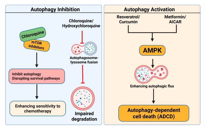
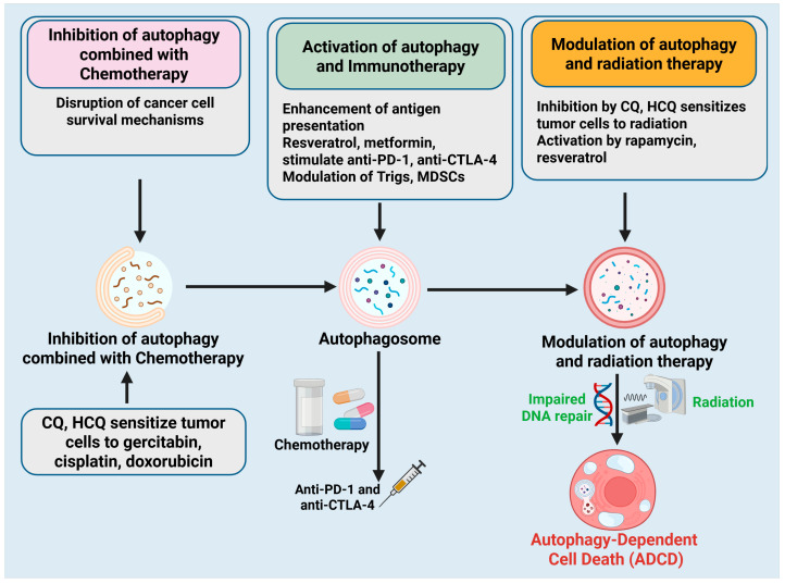
Autophagy-dependent cell death (ADCD) presents a promising but challenging therapeutic strategy in cancer treatment. Autophagy regulates cellular breakdown and stress responses, serving a dual function-either inhibiting tumorigenesis or facilitating the survival of cancer cells in advanced stages. This paradox presents both opportunities and challenges in the exploration of autophagy as a potential target for cancer treatment. In this review, we explore various pharmacological agents, including autophagy inhibitors (e.g., chloroquine, 3-MA) and activators (e.g., rapamycin, metformin), which have demonstrated effectiveness in modulating autophagy-dependent cell death (ADCD). These agents either enhance cancer cell apoptosis or sensitize tumors to conventional therapies. Combination therapies, such as the use of autophagy modulators alongside chemotherapy, immunotherapy, or radiation therapy, offer enhanced therapeutic potential by overcoming drug resistance and improving overall treatment efficacy. Nonetheless, significant challenges remain, including tumor heterogeneity, treatment resistance, and off-target effects of autophagy-targeting agents. Future progress in biomarker discovery, precision medicine, and targeted medication development will be crucial for enhancing ADCD-based methods. Although autophagy-dependent cell death presents significant potential in cancer treatment, additional studies and clinical validation are necessary to confirm its position as a conventional therapeutic approach. Therefore, this review aims to identify the existing restrictions that will facilitate the development of more effective and personalized cancer therapies, hence enhancing patient survival and outcomes.

Keywords: autophagy; autophagy-dependent cell death; cancer therapy; molecular mechanisms; tumor microenvironment.


Conflict of interest statement

The authors declare no conflicts of interest.

Figures

Figure 1
Figure 1
Molecular mechanism of autophagy. The initiation phase is governed by the ULK1 complex (comprising ULK1, ATG13, and FIP200), which is activated by AMPK and suppressed by mTORC1 signaling per food availability. Throughout elongation and maturation, the Beclin-1–VPS34 complex initiates the phagophore, whilst the ATG12-ATG5-ATG16L1 complex and lipidated LC3-II facilitate membrane extension and cargo encapsulation. Bcl-2 exerts an adverse regulatory effect on Beclin-1, hence inhibiting autophagy. During the autophagosome development phase, mature autophagosomes enclose cytoplasmic constituents, such as impaired organelles and proteins. The fusion step involves the docking of lysosomes through LAMP2, resulting in the formation of autolysosomes, where lysosomal enzymes, such as cathepsins B and D, destroy the cargo. The degradation and recycling phase facilitates the release of amino acids, lipids, and other metabolites, thus reinstating cellular equilibrium and aiding metabolic adaptability. The figure is original and was created and generated by BioRender.com, an online commercial platform.
Figure 2
Figure 2
Mechanism of autophagy-dependent cell death (ADCD). Initiation of ADCD from a healthy cell. Essential regulatory molecules, such as Beclin-1 (ATG6), ATG proteins (ATG5, ATG7, ATG12, ATG16L1), LC3 (LC3-I/LC3-II), p62/SQSTM1, and the upstream regulators mTOR and AMPK pathways, coordinate the onset and advancement of autophagy. In reaction to cellular stress, autophagosomes are formed and subsequently merge with lysosomes to create autolysosomes. Continuous autophagic activity causes cytoplasmic vacuolation and the breakdown of vital cellular components, ultimately culminating in ADCD. The function of excessive or dysregulated autophagy in facilitating non-apoptotic, planned cell death marked by significant vacuolization and organelle degradation. The figure was modified and created using the BioRender.com online commercial platform.
Figure 3
Figure 3
Interplay between autophagy-dependent cell death and other pathways. Modulating autophagy may serve as a potential therapeutic strategy in cancer treatment by influencing apoptosis, necroptosis, and ferroptosis. Autophagy modulates apoptosis by degrading anti-apoptotic proteins, including Mcl-1 and c-FLIP, hence enhancing apoptotic sensitivity. Conversely, apoptosis inhibits autophagy by caspase-3/8-mediated cleavage of Beclin-1, therefore favoring apoptotic cell death. Chemotherapeutic agents, such as doxorubicin and cisplatin, promote autophagy, thereby enhancing apoptosis in cancer treatment. Autophagy suppresses necroptosis by modulating RIPK1 (RIPK1ophagy) and phosphorylated MLKL, hence obstructing necroptotic execution. Autophagy inhibition leads to the accumulation of RIPK1, triggering necroptotic cell death. Targeting autophagy-related genes, such as ATG5 or Beclin-1, enhances the susceptibility of tumor cells to necroptosis. Ferritinophagy, facilitated by NCOA4, induces ferroptosis by liberating iron, enhancing ROS generation, and promoting lipid peroxidation. Inhibiting autophagy with chloroquine or bafilomycin A1 obstructs ferritinophagy, thereby averting ferroptosis. Conversely, ferroptosis inducers like erastin and RSL3 promote ferritinophagy, hence intensifying ferroptotic cell death. The figure was modified and created using the BioRender.com online commercial platform.
Figure 4
Figure 4
The dual role of autophagy-dependent cell death in cancer progression and therapy resistance. (Left Panel) (Tumor Suppressor Role): In the initial stages of cancer development, autophagy facilitates the removal of damaged organelles through mitophagy, decreases reactive oxygen species (ROS), and preserves genomic stability. It promotes the degradation of misfolded protein aggregates, activates tumor suppressor pathways (TP53 and PTEN), and suppresses chronic inflammation by reducing pro-inflammatory cytokines (IL-6, TNF-α), thereby inhibiting malignant transformation. (Right Panel) The right panel illustrates the role of autophagy as a tumor enhancer in advanced cancer, facilitating tumor cell survival during stress conditions such as hypoxia and nutrient deprivation, primarily through HIF-1α signaling. It improves resistance to chemotherapy and radiation by maintaining cellular integrity and inhibiting apoptosis. Autophagy sustains quiescent tumor cells in a dormant state, thereby facilitating recurrence after treatment. The shift in autophagy from a suppressive to a promotive role takes place throughout the cancer progression timeline, underscoring its complexity as a therapeutic target. The figure was modified and created using the BioRender.com online commercial platform.
Figure 5
Figure 5
Pharmacological modulation of Autophagy-dependent cell death in cancer therapy. (Left panel): Autophagy Inhibition: Chloroquine (CQ) and hydroxychloroquine (HCQ) inhibit the fusion of autophagosomes and lysosomes by increasing lysosomal pH, resulting in compromised degradation of cellular components. mTOR inhibitors have been demonstrated to indirectly inhibit autophagy or act in conjunction with CQ, thereby disrupting survival pathways and enhancing sensitivity to chemotherapy. (Right panel): Autophagy Activation: Resveratrol, curcumin, metformin, and AICAR stimulate the AMPK signaling pathway, enhancing autophagic flux. This results in ADCD, which is especially advantageous in apoptosis-resistant cancer cells. The activation of AMPK through metabolic stress or pharmacological agents facilitates the formation of autophagosomes and the degradation of oncogenic proteins. The figure was modified and created using the BioRender.com online commercial platform.
Figure 6
Figure 6
Combination strategies using Autophagy-dependent cell death modulation in cancer therapy. Three principal combinatorial techniques that employ autophagy manipulation to improve cancer treatment efficacy. (Left panel): Autophagy inhibition in conjunction with chemotherapy. Treatments like CQ and HCQ interfere with autophagy-dependent survival mechanisms in cancer cells, rendering them more susceptible to chemotherapeutic treatments such as gemcitabine, cisplatin, and doxorubicin. (Middle panel): The activation of autophagy augments immunotherapy. Autophagy inducers (e.g., resveratrol, metformin) enhance antigen presentation and immune cell activation, hence augmenting the efficacy of immune checkpoint inhibitors such as anti-PD-1 and anti-CTLA-4. Autophagy reprograms immunosuppressive cells, such as Tregs and MDSCs, into pro-inflammatory phenotypes. (Right panel): Regulation of autophagy in conjunction with radiation therapy. CQ and HCQ augment radiosensitivity by disrupting DNA repair processes, whereas compounds such as rapamycin and resveratrol may facilitate ADCD, hence amplifying the harmful effects of radiation. The figure was modified and created using the BioRender.com online commercial platform.

Similar articles

References

    1. Tobore T.O. On the need for the development of a cancer early detection, diagnostic, prognosis, and treatment response system. Future Sci. OA. 2020;6:FSO439. doi: 10.2144/fsoa-2019-0028. - DOI - PMC - PubMed
    1. Kaur R., Bhardwaj A., Gupta S. Cancer treatment therapies: Traditional to modern approaches to combat cancers. Mol. Biol. Rep. 2023;50:9663–9676. doi: 10.1007/s11033-023-08809-3. - DOI - PubMed
    1. Gu Y., Yang R., Zhang Y., Guo M., Takehiro K., Zhan M., Yang L., Wang H. Molecular mechanisms and therapeutic strategies in overcoming chemotherapy resistance in cancer. Mol. Biomed. 2025;6:2. doi: 10.1186/s43556-024-00239-2. - DOI - PMC - PubMed
    1. Bialik S., Dasari S.K., Kimchi A. Autophagy-dependent cell death–where, how and why a cell eats itself to death. J. Cell Sci. 2018;131:jcs215152. doi: 10.1242/jcs.215152. - DOI - PubMed
    1. Rahman M.A., Saikat A.S.M., Rahman M.S., Islam M., Parvez M.A.K., Kim B. Recent update and drug target in molecular and pharmacological insights into autophagy modulation in cancer treatment and future progress. Cells. 2023;12:458. doi: 10.3390/cells12030458. - DOI - PMC - PubMed

Substances

LinkOut - more resources