

Maintenance and Reversibility of Paroxysmal Atrial Fibrillation in JDP2 Overexpressing Mice
- PMID: 40710332
- PMCID: PMC12293123
- DOI: 10.3390/cells14141079
Maintenance and Reversibility of Paroxysmal Atrial Fibrillation in JDP2 Overexpressing Mice
Abstract
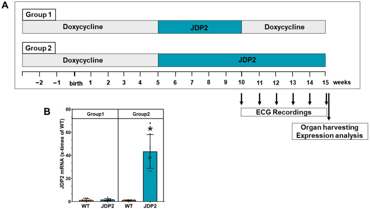
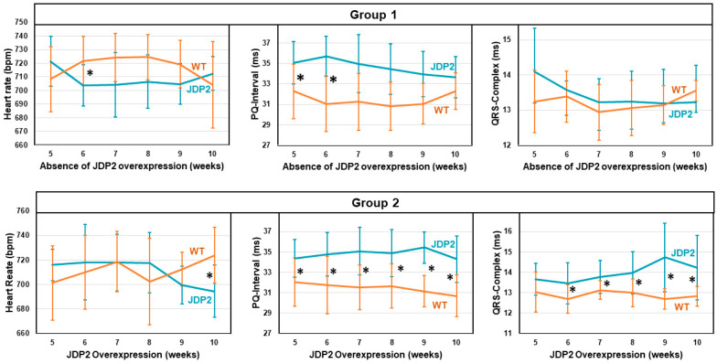
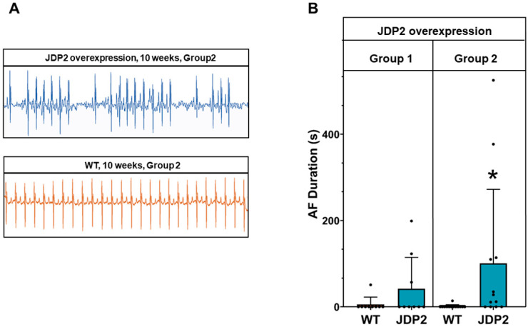
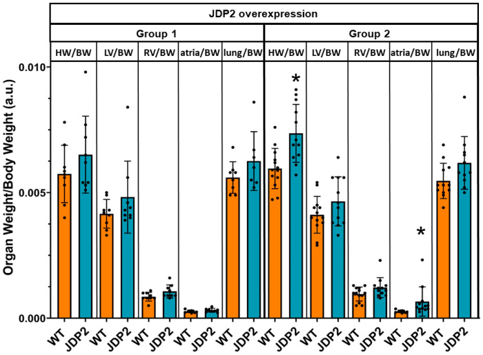
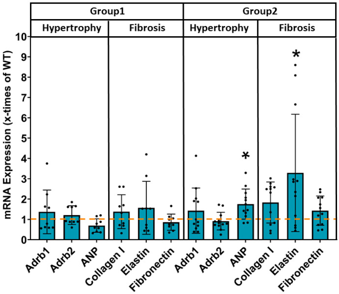
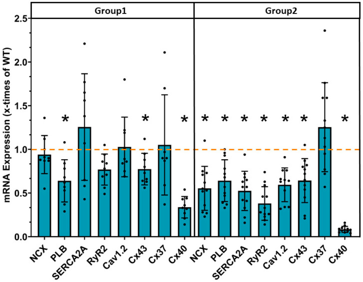
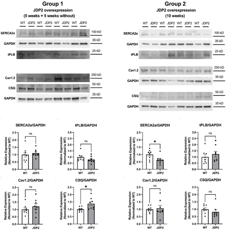
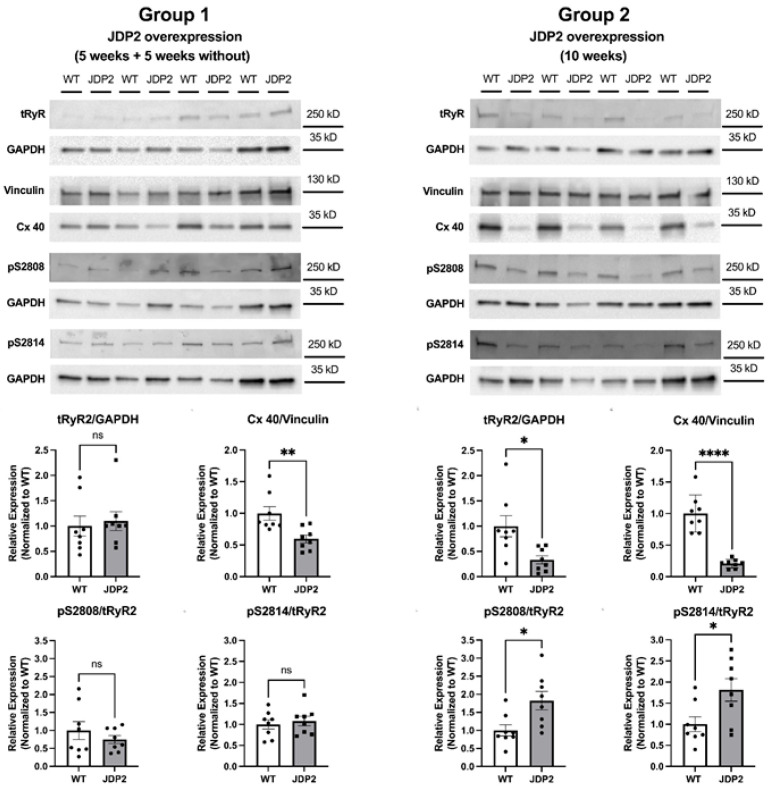
Heart-specific overexpression of transcriptional regulator JDP2 (jun dimerization protein 2) for 5 weeks provokes paroxysmal atrial fibrillation (AF) in mice. We now investigated whether AF and atrial remodeling will be reversible upon termination of JDP2 overexpression, and whether paroxysmal AF converts to permanent AF in the presence of maintained JDP2 overexpression. Cardiac-specific JDP2 overexpression for 5 weeks, resulting in paroxysmal AF, was either continued or repressed via a tet-off system for another 5 weeks. ECGs were recorded weekly. Thereafter, heart and lung weights, and atrial mRNA and protein expression were determined. Extending JDP2 overexpression did not aggravate the AF phenotype, still paroxysmal AF, prolongation of PQ intervals, and atrial hypertrophy were present. This phenotype was completely reversible upon cessation of JDP2 overexpression. A massive downregulation of connexin40 and calcium handling proteins, including SERCA2a, calsequestrin, and ryanodine receptor, was observed in atria after prolonged JDP2 overexpression. In conclusion, atrial remodeling and paroxysmal AF under JDP2 overexpression are not sufficient to maintain or aggravate AF in the absence of JDP2. The comparison of the two groups indicates that the downregulation of calcium proteins and connexins is an important factor in the maintenance of the disease.
Keywords: ECG; JDP2; SERCA2a; atrial hypertrophy; calcium; connexin; paroxysmal atrial fibrillation; ryanodine receptor.
Conflict of interest statement
The authors declare no conflicts of interest.
Figures
Similar articles
-
Structural, Pro-Inflammatory and Calcium Handling Remodeling Underlies Spontaneous Onset of Paroxysmal Atrial Fibrillation in JDP2-Overexpressing Mice.Int J Mol Sci. 2020 Nov 30;21(23):9095. doi: 10.3390/ijms21239095. Int J Mol Sci. 2020. PMID: 33265909 Free PMC article.
-
Paradoxical SERCA2a Dysregulation Contributes to Atrial Fibrillation in a Model of Diet-Induced Obesity.Int J Mol Sci. 2025 Jun 11;26(12):5603. doi: 10.3390/ijms26125603. Int J Mol Sci. 2025. PMID: 40565066 Free PMC article.
-
Atrial remodelling and atrial fibrillation self-sustaining: the role of circulating circDGCR8.Cardiovasc Res. 2025 Jul 8;121(7):1036-1051. doi: 10.1093/cvr/cvaf060. Cardiovasc Res. 2025. PMID: 40336343
-
Mechanisms of stretch-induced electro-anatomical remodeling and atrial arrhythmogenesis.J Mol Cell Cardiol. 2024 Aug;193:11-24. doi: 10.1016/j.yjmcc.2024.05.011. Epub 2024 May 24. J Mol Cell Cardiol. 2024. PMID: 38797242 Free PMC article. Review.
-
Curative catheter ablation in atrial fibrillation and typical atrial flutter: systematic review and economic evaluation.Health Technol Assess. 2008 Nov;12(34):iii-iv, xi-xiii, 1-198. doi: 10.3310/hta12340. Health Technol Assess. 2008. PMID: 19036232
References
-
- Joglar J.A., Chung M.K., Armbruster A.L., Benjamin E.J., Chyou J.Y., Cronin E.M., Deswal A., Eckhardt L.L., Goldberger Z.D., Gopinathannair R., et al. 2023 ACC/AHA/ACCP/HRS Guideline for the Diagnosis and Management of Atrial Fibrillation: A Report of the American College of Cardiology/American Heart Association Joint Committee on Clinical Practice Guidelines. Circulation. 2024;149:e1–e156. doi: 10.1161/CIR.0000000000001193. - DOI - PMC - PubMed
-
- Aguilar M., Macle L., Deyell M.W., Yao R., Hawkins N.M., Khairy P., Andrade J.G. Influence of Monitoring Strategy on Assessment of Ablation Success and Postablation Atrial Fibrillation Burden Assessment: Implications for Practice and Clinical Trial Design. Circulation. 2022;145:21–30. doi: 10.1161/CIRCULATIONAHA.121.056109. - DOI - PubMed
MeSH terms
Substances
LinkOut - more resources
Full Text Sources
Medical
Miscellaneous
