

Microvascularization of the Vocal Folds: Molecular Architecture, Functional Insights, and Personalized Research Perspectives
- PMID: 40710410
- PMCID: PMC12299232
- DOI: 10.3390/jpm15070293
Microvascularization of the Vocal Folds: Molecular Architecture, Functional Insights, and Personalized Research Perspectives
Abstract
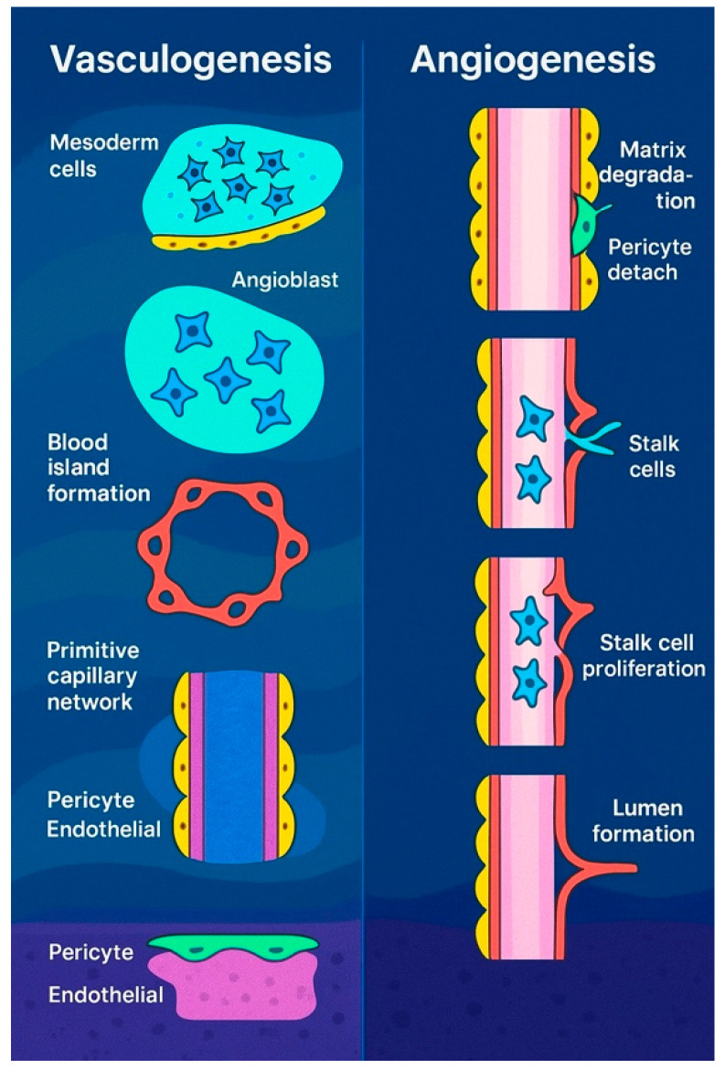
Introduction: The vascular architecture of the vocal folds plays a critical role in sustaining the dynamic demands of phonation. Disruptions in this microvascular system are linked to various pathological conditions, including Reinke's edema, hemorrhage, and laryngeal carcinoma. This review explores the structural and functional components of vocal fold microvascularization, with emphasis on pericytes, endothelial interactions, and neurovascular regulation. Materials and Methods: A systematic review of the literature was conducted using databases such as PubMed, Scopus, Web of Science, and Embase. Keywords included "pericytes", "Reinke's edema", and "vocal fold microvascularization". Selected studies were peer-reviewed and met criteria for methodological quality and relevance to laryngeal microvascular physiology and pathology. Results: The vocal fold vasculature is organized in a parallel, tree-like pattern with distinct arterioles, capillaries, and venules. Capillaries dominate the superficial lamina propria, while transitional vessels connect to deeper arterioles surrounded by smooth muscle. Pericytes, present from birth, form tight associations with endothelial cells and contribute to capillary stability, vessel remodeling, and mechanical protection during vibration. Their thick cytoplasmic processes suggest a unique adaptation to the biomechanical stress of phonation. Arteriovenous anastomoses regulate perfusion by shunting blood according to functional demand. Furthermore, neurovascular control is mediated by noradrenergic fibers and neuropeptides such as VIP and CGRP, modulating vascular tone and glandular secretion. The limited lymphatic presence in the vocal fold mucosa contributes to edema accumulation while also restricting carcinoma spread, offering both therapeutic challenges and advantages. Conclusions: A deeper understanding of vocal fold microvascularization enhances clinical approaches to voice disorders and laryngeal disease, offering new perspectives for targeted therapies and regenerative strategies.
Keywords: Reinke’s edema; capillary permeability; endothelial cells; microvascularization; pericytes; personalized vascular signaling; vocal folds.
Conflict of interest statement
The authors declare no conflicts of interest.
Figures
Similar articles
-
Laryngeal Applications of Platelet Rich Plasma and Platelet Poor Plasma: A Systematic Review.J Voice. 2024 Jan;38(1):248.e1-248.e13. doi: 10.1016/j.jvoice.2021.07.007. Epub 2021 Aug 10. J Voice. 2024. PMID: 34384663
-
Magnetic resonance perfusion for differentiating low-grade from high-grade gliomas at first presentation.Cochrane Database Syst Rev. 2018 Jan 22;1(1):CD011551. doi: 10.1002/14651858.CD011551.pub2. Cochrane Database Syst Rev. 2018. PMID: 29357120 Free PMC article.
-
Musical and vocal interventions to improve neurodevelopmental outcomes for preterm infants.Cochrane Database Syst Rev. 2023 Sep 7;9(9):CD013472. doi: 10.1002/14651858.CD013472.pub2. Cochrane Database Syst Rev. 2023. PMID: 37675934 Free PMC article.
-
Effects of voice therapy in children with vocal fold nodules: A systematic review.Int J Lang Commun Disord. 2022 Nov;57(6):1160-1193. doi: 10.1111/1460-6984.12754. Epub 2022 Jun 27. Int J Lang Commun Disord. 2022. PMID: 35758272
-
The experience of adults who choose watchful waiting or active surveillance as an approach to medical treatment: a qualitative systematic review.JBI Database System Rev Implement Rep. 2016 Feb;14(2):174-255. doi: 10.11124/jbisrir-2016-2270. JBI Database System Rev Implement Rep. 2016. PMID: 27536798
References
-
- Rzepakowska A., Żurek M., Grzybowski J., Pihowicz P., Górnicka B., Osuch-Wójcikiewicz E., Niemczyk K. Correlation of narrow band imaging vascular patterns with immunohistological microvessel density in vocal fold lesions. Braz. J. Otorhinolaryngol. 2021;87:137–144. doi: 10.1016/j.bjorl.2019.07.009. - DOI - PMC - PubMed
-
- Wang Y. Seeing the future of histotechnology through its history. J. Histotechnol. 2018;41:135–136. doi: 10.1080/01478885.2018.1527590. - DOI
-
- Thomas A.M., Banerjee A.K. The History of Radiology. OUP Oxford; Oxford, UK: 2013.
-
- Hînganu M.V., Cozma R.S., Ciochina P., Scutariu I.A., Asimionoaiei-Simionescu C., Hînganu D. The morphometry of the laryngeal phonatory system—Base of the anatomical study of the voice aptitudes. Rom. J. Morphol. Embryol. 2017;58:1365–1369. - PubMed
Publication types
LinkOut - more resources
Full Text Sources
Research Materials
Miscellaneous
