. 2025 Jul 2;11(7):514.
doi: 10.3390/gels11070514.

MOFs-Combining Fully Synthetic Injectable Hydrogel Scaffolds Exhibiting Higher Skeletal Muscle Regenerative Efficiency than Matrigel

Affiliations

MOFs-Combining Fully Synthetic Injectable Hydrogel Scaffolds Exhibiting Higher Skeletal Muscle Regenerative Efficiency than Matrigel

Sobuj Shahidul Islam et al. Gels. .

Abstract

Due to its sarcoma-derived origin and the associated carcinogenic risks, as well as its lack of tissue-specific extracellular matrix biochemical cues, the use of the injectable gel scaffold Matrigel is generally restricted to research applications. Therefore, the development of new fully synthetic injectable gel scaffolds that exhibit performance comparable to Matrigel is a high priority. In this study, we developed a novel fully synthetic injectable gel scaffold by combining a biodegradable PLGA-PEG-PLGA copolymer, clay nanoparticle LAPONITE®, and L-arginine-loaded metal-organic frameworks (NU-1000) at the nano level. An aqueous solution of the developed hybrid scaffold (PLGA-PEG-PLGA/LAPONITE®/L-Arg@NU-1000) exhibited rapid sol-gel transition at body temperature following simple injection and formed a continuous bulk-sized gel, demonstrating good injectability. Long-term sustained slow release of L-arginine from the resultant gels can be achieved because NU-1000 is a suitable reservoir for L-arginine. PLGA-PEG-PLGA/LAPONITE®/L-Arg@NU-1000 hybrid gels exhibited good compatibility with and promoted the growth of human skeletal muscle satellite cells. Importantly, in vivo experiments using skeletal muscle injury model mice demonstrated that the tissue regeneration efficiency of PLGA-PEG-PLGA/LAPONITE®/L-Arg@NU-1000 gels is higher than that of Matrigel. Specifically, we judged the higher tissue regeneration efficacy of our gels by histological analysis, including MYH3 immunofluorescent staining, H&E staining, and Masson's trichrome staining. Taken together, these data suggest that novel hybrid hydrogels could serve as injectable hydrogel scaffolds for in vivo tissue engineering and ultimately replace Matrigel.

Keywords: MOFs; injectable hydrogels; scaffolds; skeletal muscle tissue; tissue engineering.


Conflict of interest statement

The authors declare that the commercial products LAPONITE® and PLGA-PEG-PLGA/LAPONITE®/L-Arg@NU-1000 do not involve any conflicts of interest.

Figures

Figure 1
Figure 1
Schematic illustration of the structure and function of the developed PLGA-PEG-PLGA/LAPONITE®/L-Arg@NU-1000 injectable hydrogels for the promotion of skeletal muscle tissue regeneration.
Figure 2
Figure 2
(A) Temperature-dependent rheological properties of the PLGA-PEG-PLGA/ LAPONITE®/L-Arg@NU-1000 hydrogels and the PLGA-PEG-PLGA/LAPONITE® hydrogel. (B) Time-dependent rheological properties of the PLGA-PEG-PLGA/LAPONITE®/L-Arg@NU-1000 hydrogels and the control PLGA-PEG-PLGA/LAPONITE® hydrogel measured at 37 °C.
Figure 3
Figure 3
(A) Representative images of the injectability of the PLGA-PEG-PLGA/LAPONITE® gel (left) and the PLGA-PEG-PLGA/LAPONITE®/L-Arg@NU-1000 gel (right) tested at 37 °C. (B) Representative image of the self-standing PLGA-PEG-PLGA/LAPONITE®/L-Arg@NU-1000 gel at 37 °C fabricated by simple injection.
Figure 4
Figure 4
Release profiles of L-arginine from the PLGA-PEG-PLGA/LAPONITE®/L-Arg@NU-1000 gels with different amounts of L-arginine loading and the control PLGA-PEG-PLGA/LAPONITE® gel.
Figure 5
Figure 5
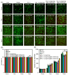
(A) Representative fluorescence microscopy images of the HsKMSCs cultured for 1, 3, and 7 days in the presence of the PLGA-PEG-PLGA/LAPONITE®/L-Arg@NU-1000 gels, the control PLGA-PEG-PLGA/LAPONITE® gel, and Matrigel. Scale bars: 100 μm. (B) Percent quantification from the proliferation of HsKMSC culture. (C) Proliferation of the cultured HsKMSCs in the presence of the PLGA-PEG-PLGA/LAPONITE®/L-Arg@NU-1000 gels, the control PLGA-PEG-PLGA/LAPONITE® gel, and Matrigel. The p-values are indicated as follows: * p < 0.05.
Figure 6
Figure 6
(A) Representative fluorescence microscopy images of HsKMSCs stained with phalloidin and DAPI, cultured for 1, 3, and 7 days in the presence of the PLGA-PEG-PLGA/LAPONITE®/L-Arg@NU-1000 gels, the control PLGA-PEG-PLGA/LAPONITE® gel, and Matrigel. Scale bars: 50 μm. (B) Average width of myotubes detected in the cultured HsKMSCs in the presence of the PLGA-PEG-PLGA/LAPONITE®/L-Arg@NU-1000 gels, the control PLGA-PEG-PLGA/LAPONITE® gel, and Matrigel. (C) Average length of myotubes detected in the cultured HsKMSCs in the presence of the PLGA-PEG-PLGA/LAPONITE®/L-Arg@NU-1000 gels, the control PLGA-PEG-PLGA/LAPONITE® gel, and Matrigel. The p-values are indicated as follows: * p < 0.05; ** p < 0.01; *** p < 0.001.
Figure 7
Figure 7
Representative images of injured and reconstructed skeletal muscle tissues after treatments with the PLGA-PEG-PLGA/LAPONITE®/L-Arg@NU-1000 gels and the control PLGA-PEG-PLGA/LAPONITE® gels for 7 and 28 days.
Figure 8
Figure 8
Representative images of injured and reconstructed skeletal muscle tissues after treatments with Matrigel for 35 days.
Figure 9
Figure 9
(A) Representative fluorescence microscopy images of reconstructed skeletal muscle tissue sections stained with CD31 after 7 and 28 days with treatments by the PLGA-PEG-PLGA/LAPONITE®/L-Arg@NU-1000 gels and the control PLGA-PEG-PLGA/LAPONITE® gels. Scale bars: 100 μm. (B) Average number of CD31-positive blood vessels in the reconstructed skeletal muscle tissue section after 7 and 28 days with treatments by the PLGA-PEG-PLGA/LAPONITE®/L-Arg@NU-1000 gels and the control PLGA-PEG-PLGA/LAPONITE® gels. The p-values are indicated as follows: * p < 0.05.
Figure 10
Figure 10
(A) Representative fluorescence microscopy images of reconstructed skeletal muscle tissue sections stained with CD31 after 35 days with treatments by Matrigel. Scale bars: 100 μm. (B) Average number of CD31-positive blood vessels in the reconstructed skeletal muscle tissue section with treatments by the PLGA-PEG-PLGA/LAPONITE®/L-Arg@NU-1000 gels and Matrigel. The p-values are indicated as follows: * p < 0.05. Significant changes were identified by comparing each with mean 2% ± SD.
Figure 11
Figure 11
(A) Representative fluorescence microscopy images of reconstructed skeletal muscle tissue sections stained with MYH3 after 7 and 28 days with treatments by the PLGA-PEG-PLGA/LAPONITE®/L-Arg@NU-1000 gels and the control PLGA-PEG-PLGA/LAPONITE® gels. Scale bars: 100 μm. (B) Average number of MYH3-positive myofibers in the reconstructed skeletal muscle tissue section after 7 and 28 days with treatments by the PLGA-PEG-PLGA/LAPONITE®/L-Arg@NU-1000 gels and the control PLGA-PEG-PLGA/LAPONITE® gels. The p-values are indicated as follows: * p < 0.05.
Figure 12
Figure 12
(A) Representative fluorescence microscopy images of reconstructed skeletal muscle tissue sections stained with MYH3 after 35 days with treatments by Matrigel. Scale bars: 100 μm. (B) Average number of CD31-positive myofibers in the reconstructed skeletal muscle tissue section with treatments by the PLGA-PEG-PLGA/LAPONITE®/L-Arg@NU-1000 gels and Matrigel. The p-values are indicated as follows: * p < 0.05.
Figure 13
Figure 13
(A) Representative microscopy images of reconstructed skeletal muscle tissue sections stained with H&E after 7 and 28 days with treatments by the PLGA-PEG-PLGA/LAPONITE®/L-Arg@NU-1000 gels and the control PLGA-PEG-PLGA/LAPONITE® gels. Scale bars: 300 μm. (B) Reconstructed area (%) of skeletal muscle tissue sections after 7 and 28 days with treatments by the PLGA-PEG-PLGA/LAPONITE®/L-Arg@NU-1000 gels and the control PLGA-PEG-PLGA/LAPONITE® gels. (C) Area of the reconstructed skeletal muscle in the whole reconstructed tissues after 7 and 28 days with treatments by the PLGA-PEG-PLGA/LAPONITE®/L-Arg@NU-1000 gels and the control PLGA-PEG-PLGA/LAPONITE® gels. The p-values are indicated as follows: * p < 0.05; ** p < 0.01; *** p < 0.001.
Figure 14
Figure 14
(A) Representative microscopy images of reconstructed skeletal muscle tissue sections stained with H&E after 35 days with treatment by Matrigel. Scale bars: 300 μm. (B) Reconstructed area (%) in the skeletal muscle tissue section after 35 days with treatment by Matrigel. (C) Area of the reconstructed skeletal muscle in whole reconstructed tissues with treatments by the PLGA-PEG-PLGA/LAPONITE®/L-Arg@NU-1000 gel and Matrigel. The p-values are indicated as follows: * p < 0.05.
Figure 15
Figure 15
(A) Representative microscopy images of reconstructed skeletal muscle tissue sections after Masson trichrome staining, 7 and 28 days after treatments by the PLGA-PEG-PLGA/LAPONITE®/L-Arg@NU-1000 hydrogels and the control PLGA-PEG-PLGA/LAPONITE® gels. Scale bars: 300 μm. (B) Area of fibrosis in the reconstructed skeletal muscle tissues on 7 and 28 days after treatments by the PLGA-PEG-PLGA/LAPONITE®/L-Arg@NU-1000 gels and the control PLGA-PEG-PLGA/LAPONITE® gels. * p < 0.05, ** p < 0.01, *** p < 0.001.
Figure 16
Figure 16
(A) Representative microscopy images of reconstructed skeletal muscle tissue sections after Masson trichrome staining, 35 days after treatment by Matrigel. Scale bars: 300 μm. (B) Area of fibrosis in the reconstructed skeletal muscle tissues after treatments by the PLGA-PEG-PLGA/LAPONITE®/L-Arg@NU-1000 hydrogels and Matrigel.

References

    1. Kitagawa S., Kitaura R., Noro S.I. Functional porous coordination polymers. Angew. Chem. Int. Ed. Engl. 2004;43:2334–2375. doi: 10.1002/anie.200300610. - DOI - PubMed
    1. Li H., Eddaoudi M., Keeffe M.O., Yaghi O.M. Design and synthesis of an exceptionally stable and highly porous metal-organic framework. Nature. 1999;402:276–279. doi: 10.1038/46248. - DOI
    1. He Y., Zhou W., Qian G., Chen B. Methane storage in metal-organic frameworks. Chem. Soc. Rev. 2014;43:5657–5678. doi: 10.1039/C4CS00032C. - DOI - PubMed
    1. Kitagawa S. Future porous materials. Acc. Chem. Res. 2017;50:514–516. doi: 10.1021/acs.accounts.6b00500. - DOI - PubMed
    1. Daglar H., Gulbalkan H.C., Avci G., Aksu G.O., Altundal O.F., Altintas C., Erucar L., Keskin S. Effects of metal-organic framework (MOF) database selection on the assessment of gas storage and separation potentials of MOFs. Angew. Chem. Int. Ed. Engl. 2021;60:7828–7837. doi: 10.1002/anie.202015250. - DOI - PMC - PubMed

Grants and funding

LinkOut - more resources